Transport and Respiration Sample Assignment
Task 1:
1.1. Main Components and Function of Blood
Components
Blood is the connective tissue in the human body. It mainly comprises four elements- Plasma, Platelets, Red Blood cells, and White blood cells.
Plasma
Plasma is an extracellular fluid which is a mixture of enzymes, proteins, hormones, nutrients, wastes, and gases. It helps in balancing the fluids of the body by transporting these components in different parts.
Red Blood Cells
Red blood cells contain about 40% of the whole volume of blood in a body. It is produced from the erythropoietin and has no nucleus. It changes its shape accordingly. Red blood cells have hemoglobin that helps in carrying oxygen to the lungs.
White Blood Cells
White blood cells are the one that protects the body from any kind of infection. They consist of 15 of total blood volume. White blood cells have lymphocytes and neutrophil. These blood cells help to build immunity against tumors and infected cells.
Platelets
These fragments of blood cells help in coagulation in an injured blood vessel. The blood clotting is the main function of these blood cells.
Functions
Blood is body fluid that has mainly three functions, these are Protection, regulation, and Transport. The functions of blood can be discussed as below-
Transport
- Transportation of the important nutrients, oxygen, and carbon-dioxide is done by blood
- As a carrier, blood also carries the wastes to the kidney and liver. These organs filter the wastes from the blood and keep it clean.
- It carries the hormones from their glands and transports it to the designated organs and body parts.
Protection
- It has an important role in fighting against infection. The blood carries the antibodies and cells so that the body can fight infections.
- Different pathogenic substances are destroyed by the proteins and antibodies in the blood.
- The platelets in the blood form blood clots so that body could prevent excess loss of blood from the body.
Regulation
- Blood has an important part in maintaining and regulating body temperature.
- Blood interacts with bases and acids and regulates the pH of the human body (myvmc.com, (2019).
Task 2
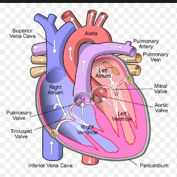
2.1 a) heart dissection is a critical operation where safety is required. There is a line in front of the heart and make a deep incision on it and the incision will be made over the top wall of a heart. After that heart dissected in two parts. There are chambers which include atrium and ventricles. There are many other parts in the heart which include valves, papillary muscle, and chordae tendineae [referred to appendix 1].
|
|
|
- b) Heart has cardiac muscle tissue is found in the heart which helps to pump the blood through the body. Other than this heart has highly dense connective tissue which helps to anchor and form valves of the heart. According to Lai et al. (2015), there are four chambers which are left ventricle, left atrium, right ventricle, and right atrium. Aorta which is the largest artery present in heart helps to carry out oxygenated blood to the body from left ventricle. Vein present is superior vena cava that helps to carry out deoxygenated blood from body to heart. Inferior vena cava brings deoxygenated blood to heart from the lower part of the body. Pulmonary artery present, that helps to carry blood, which is deoxygenated to lungs from the right ventricle. Pulmonary vein present which carries blood oxygenated to left atrium to the lung. There is a tricuspid valve that helps in the prevention of blood flow back in the right atrium (Elliott et al. 2017). Four chambers of heart help to store the blood and carry to other places with the help of arteries and veins.
Task 3
3.1. a) Coronary, systematic and pulmonary circulation
The circulatory system has the function of balancing all another system by maintaining body temperature, fighting diseases and providing correct chemical balance. The four main components that are invested in the circulatory system are blood, veins, arteries, and heart. The constant pressure from the heart and the valves help the regulation.
Pulmonary Circulation
In this circulation system, the deoxygenated blood is carried away from the right ventricle of the heart and is carried to lungs. Again the oxygenated blood is carried to the left arteries and ventricle of the heart.
Coronary Circulation
The circulation of the oxygenated blood is supplied to the heart by the coronary arteries and the deoxygenated blood is carried away from it by coronary veins.
Systemic Circulation
This portion of the cardiovascular system transports the oxygenated blood from the left ventricle of heart away through aorta where the pulmonary circulation has deposited the blood, to the best part of the body. The oxygen-depleted blood is brought back to the heart (livescience.com, 2019).
Difference
The main difference between these circulation systems lies in the Pulmonary and systemic circulation. In the Pulmonary circulation, the closed loop vessels carry the blood between lungs and heart, whereas in the systemic circulation the blood is carried between the heart and other parts of the body.
- b) The given example has an effect of the disruption of the pulmonary circulation. The blood clot blocks one or more than one arteries and the patient suffers from a stroke in this kind of embolism.
The effect of this kind of embolism may include sudden chest pain and a shortage of breath, dry cough, fall of oxygen level in the body and palpitation. These are the most common effects of this kind of stroke caused by pulmonary circulation disruption. This kind of embolism is called pulmonary embolism.
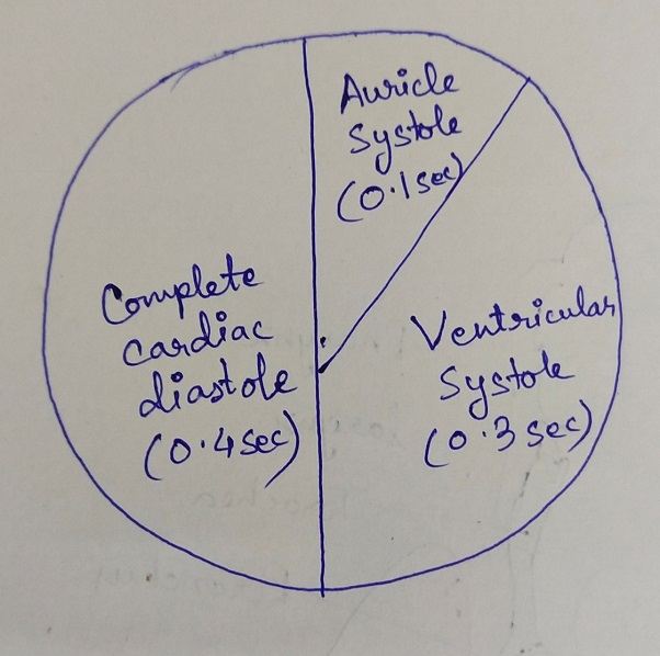
3.2. a) cardiac cycle
There are seven phases of a cardiac cycle and have a mechanism of systole and diastole. One cardiac cycle refers to one systole and one diastole.
First phase: at this stage arterial contraction happens and there is a pressure created and blood rush to ventricle with the help of mitral valve. At this time aortic valve stays closed.
Second phase: at this stage systole ventricle happens and it creates pressure which closes the mitral valve and increases pressure on valve intraventricular (Aghamohammadzadeh et al. 2015). At this stage, the first sound of the heart is produced which is S1. Due to the accumulation of blood intra atrial pressure increases.
Third phase: after the increase of pressure, the opening of aortic valve happens. Due to the contraction of ventricle intraventricular pressure gets increased. At this stage, aorta, and ventricle work as the unit chamber. At this stage left atrium receives the blood and blood accumulation in the atrium happens which create wave ‘v’.
Fourth phase: ventricle is kept on contracting however aorta pressure starts to fail. At this stage, intraventricular pressure stays increased. At this phase, the aortic valve stays open and aorta contracts to pump the blood into an arterial peripheral tree.
Fifth phase: at this stage ventricle become relaxed and aortic valve became closed. In this time isovolumetric relaxation occurred (Brunt et al. 2016). In this stage, atrium work as a blood reservoir.
Sixth stage: at this left ventricle became relaxed totally and pressure in atrium became higher than left ventricle. Therefore, mitral valve opens at this stage. Accumulated blood in atrium go to ventricle and contraction of atrium takes place.
Seventh phase: with theopening of the atrioventricular valve, blood goes to ventricle from atrium directly.

Figure 1: cardiac cycle
(Source: created by the author)
- b) congenital heart defects such as a hole in the heart disrupts cardiac cycle such as loud murmur sound can be heard. As opined by Hutchinson et al. (2016), moreover high pressure in the left heart can be seen which force the blood through VSD to the right heart. At the same time, oxygenated and deoxygenated blood gets mixed and blood never gets purified in the heart. Therefore, deoxygenated blood passes through all over the body.
3.3) a) cardiac output and blood pressure
The amount of blood gets pumped by the heart per minute is known as cardiac output. Heart rate and stroke volume product are known as cardiac output. Heart rate refers to heartbeat per minute. The volume of stroke refers to blood amount which pumped per minute. According to Abdelkader et al. (2016), cardiac output is different to different people depending upon the size of that person. It increases and decreased as per the exercise is done by a person or at rest. As at rest, the pumping of the blood of an adult is 3-4 litres and during exercise, it increases to 3-4 times as the body requires more oxygen.
Blood pressure is force measurement of heart which determines how much blood gets pumped all over the body with the help of the heart. There are main two pressures which are diastolic pressure and systolic pressure (Chiesa et al. 2016). During diastolic pressure, between beats hearts stay at rest condition. At systolic pressure, the heart pushes the blood all over the body. Normal blood pressure of an adult is 120/80.
- b) at the same range of right atrial pressure, at rest stage cardiac output is relatively lower which 6L/min. On the other hand, cardiac output is relatively higher during exercise which is around 14L/min. Therefore, it suggests that during exercise body requires more oxygen and stroke volume became increased and more blood pumps from the heart to all over the body. On the other hand, at rest requirement of oxygen is less therefore cardiac output also became less (Lin et al. 2018). Moreover, at rest blood pumps out all over the body from heart relatively lower rate than exercise stage. At rest, heart eats also stay relatively low.
Task 4
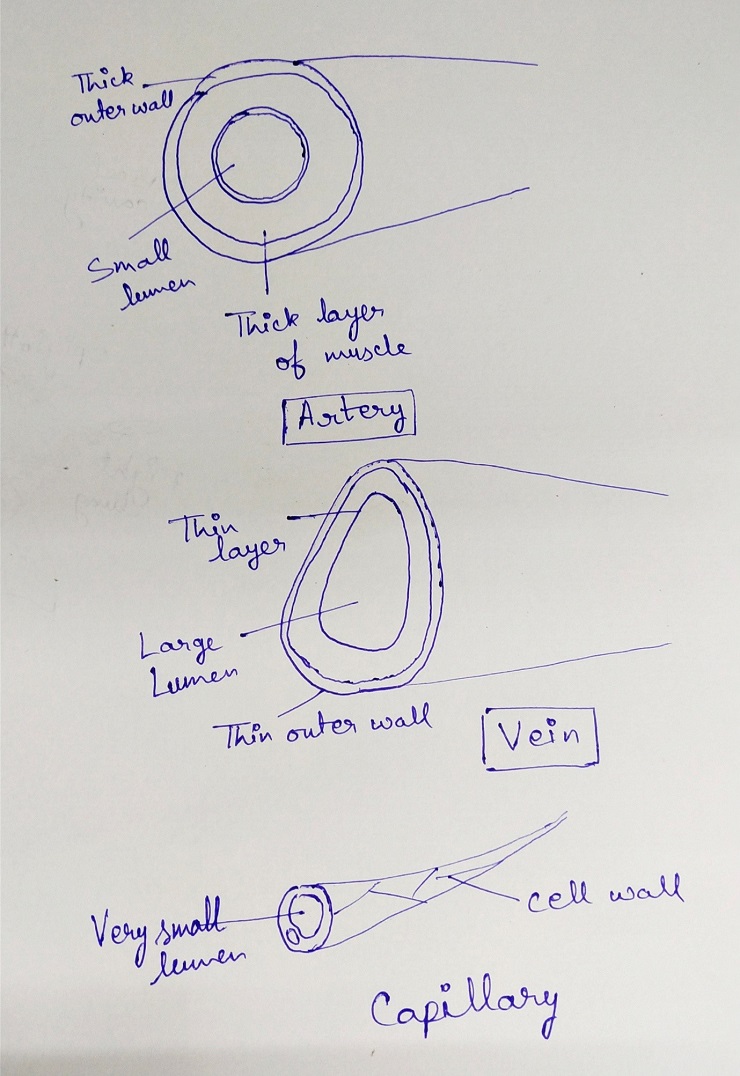
4.1 a) Structure of arteries, capillaries, and veins in regard to their functions
Arteries, capillaries and the veins have the structure that maintains a different kind of function in the human respiratory system.
Arteries
There is a thick layer in the outer part of arteries that is consisted of collagen and fibres. This helps in avoiding the leakages and bulges. The high pressure in the respiratory process is held by the thick wall of arteries.
The blood is pump after every contraction in the heart through the elastic and muscle fibres that constitute the thick layer in arteries. The high pressures inside arteries are maintained by narrow lumen.
Veins
The main components of the veins are made up by thin layers of circular muscles and fibres. The blood flow is not there in the pulses, that is why the walls of the vein do not pump blood.
The thin walls help them to become flat whenever it feels the pressure from the nearby muscles. The blood is pushed forward in the direction of heart by this process. The thin layers do not burst as there is low pressure in the veins. An outer layer of the vein is constituted on longitudinal collagen and fibres. The low pressure of the veins is accommodated by the wide lumen. Unlike arteries, veins have valves that prevent blood pooling.
Capillaries
The wall of the capillaries is only built will one layer of cells. The diffusion is the main work of capillaries and the small shape of it make the diffusion rapid. Capillaries exchange the gas and materials with the other surrounding tissues in the body. The tissue fluids that are leaked from the thin wall of capillaries help in transporting the materials. The pores of the capillaries help in phagocytes passing.
The narrow structure of the capillary lumen can fit the small space and that helps in increasing the surface areas for diffusion (mytutor.co.uk, 2019).

Figure 2: vein, artery and capillary
(Source: created by the author)
- b) In the given case the arterial vessels are haemorrhaging. This kind of haemorrhage is severe with respect to the other types of blood vessels. The arteries are the means of transporting the oxygenated blood from the heart to the other parts of the body. The haemorrhage in arteries can block the whole process and the patient can face paralytic condition for the lack of oxygen in the brain and other body parts.
Task 5
5.1)
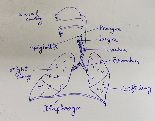
- b) Nasal cavity
The respiratory system opening is the nasal cavity. It is also the main opening which is external. It is the of the airway in the body. It is the tract of the respiratory system through which air can pass. It filters, moisturizes and warms the air that enters the body before it gets to the lung ( Duffelse et al. 2017). The mucus and the hair lining trap dust, pollen, mold, and other particles. The air that exits the nasal cavity returns its warmth and moisture.
Pharynx
The throat is also known as the pharynx. It consists of three different regions, such as oropharynx, nasopharynx, and laryngopharynx. The nasopharynx is the region that is superior. The air which is inhaled passes into the nasopharynx through the oropharynx. Then it finally descends to the laryngopharynx. The main function of the pharynx is to swallow food. The main function of the epiglottis is to help the air pass to the trachea. When the food is swallowed, the trachea is covered by the epiglottis. This prevents the food from entering into the epiglottis.
Larynx
The larynx is the voice box. It connects the laryngopharynx to the trachea. It is the airway's short section. The interior of the neck is the location of the larynx. The larynx is made up of multiple cartilage structures (Hamilton et al. 2012). The larynx is also made of structures defined as the voice fold. It allows the body to produce the sound of singing and speech. The tension and speed of vibration enable the fold of voice to change pitch that is produced.
- Trachea
The trachea is made up of cartilage and comes from the larynx and goes to bronchi. It is large air tubes and almost 4 inches long and 1 inch in diameter. The main function of the trachea is to act as a passageway. Through passageway, air passes to the main respiratory organ which is bronchi from voice box after that bronchus enters the lungs.
- The pleural cavity and pleural membrane
Within the pleural cavity, each lung is placed. On the other hand, pleural membrane cover lung's outer layer. It also helps to line inside chest wall. There are two membranes in the pleural membrane which are visceral pleura and parietal pleura (Imai et al. 2015). The pleural cavity is filled with a serious fluid which makes the lung easy to move within the thoracic cavity. The fluid also helps in keeping lung in close contact with thorax wall and also provides surface tension.
- Lung
- I) the singular form of bronchi is known as bronchus. Each bronchus consists of cartilage, smooth muscle and also mucosal lining. Bronchi help in the process of gas exchange. It helps to carry the blood to lung and carbon dioxide leaves the lung and help in exchange of gas. In the respiratory system, bronchi are known as the conducting zone.
- II) Bronchioles help to pass the air with the help of mouth or nose to the air sacs which are alveoli. There are capillaries or tiny blood vessels are present which surrounds alveoli (Lai et al. 2015).
III) Alveoli are the essential part of the respiratory system which helps in exchange of carbon dioxide and oxygen molecule in the bloodstream. All of them stay as cluster and balloon shaped at the end of the tree of respiration.
7) intercostal muscle
Help in the expansion of the chest cavity and also help quiet as well as forced inhalation. However, intercostal muscle helps in exhalation forcefully (Elliott et al. 2017).
8) Diaphragm
The diaphragm is located below the lungs and work as respiration's major muscle. It a dome-shaped muscle and it contracts in rhythm and constantly.

Figure 3: respiratory organs
(Source: created by the author)
Task 6
6.1 Stages of Pulmonary ventilation and mechanism behind it
The pulmonary ventilation is also known as breathing has two parts- inhalation and exhalation. Inhalation is the process of breathing where the air flowed into the lungs in the time of inspiration. Exhalation, on the other hand, is the process where the air is carried out from the lungs. The flow of the air is the result of pressure differences in the atmosphere and gases inside the lungs.
Breathing or pulmonary ventilation have three different types of pressure-
- Atmospheric pressure
- Intrapulmonary pressure
- Intrapleural pressure
The common law that works behind the flow of gas is that gas has the tendency to flow from the high-pressure region to the low-pressure region. Ventilation is the result of muscular movement and elastic tissue recoiling that makes the change in pressure happen.
The pressure outside the body is the atmospheric pressure and the other kind of pressure is the intraocular pressure. This kind of pressure acts in the inner part of alveoli of the lungs.
Inspiration
Inspiration method is the inhalation of the air in breathing. In the inspiration method, contraction of the diaphragm occurred and the volume of the thoracic cavity is increased. The intra alveolar pressure is decreased and the air flows in the lungs.
Expiration
The process of letting air out in the breathing process is called expiration. In this process, the tissue elastic recoil and diaphragm relaxation decrease the volume of thoracic. Intra Alveolar pressure is increased and the air is pushed out of the lungs (training.seer.cancer.gov,2019).
6.2) non steroid anti inflammatory medicines helps to deal with chest pains which disrupt pulmonary ventilation. It causes a collateral damage to chest which resulted in severe disruption of pulmonary ventilation.
.
Reference list
Book
Lai, W.W., Mertens, L.L., Cohen, M.S. and Geva, T., 2015. Echocardiography in pediatric and congenital heart disease: from fetus to adult. John Wiley & Sons.
Journal
Elliott, L., Sharp, K., Alfaro-Almagro, F., Douaud, G., Miller, K., Marchini, J. and Smith, S., 2017. The genetic basis of human brain structure and function: 1,262 genome-wide associations found from 3,144 GWAS of multimodal brain imaging phenotypes from 9,707 UK Biobank participants. BioRxiv, p.178806.
Aghamohammadzadeh, R., Unwin, R.D., Greenstein, A.S. and Heagerty, A.M., 2015. Effects of obesity on perivascular adipose tissue vasorelaxant function: nitric oxide, inflammation and elevated systemic blood pressure. Journal of vascular research, 52(5), pp.299-305.
Brunt, V.E., Howard, M.J., Francisco, M.A., Ely, B.R. and Minson, C.T., 2016. Passive heat therapy improves endothelial function, arterial stiffness and blood pressure in sedentary humans. The Journal of physiology, 594(18), pp.5329-5342.
Hutchinson, J.C., Arthurs, O.J., Ashworth, M.T., Ramsey, A.T., Mifsud, W., Lombardi, C.M. and Sebire, N.J., 2016. Clinical utility of postmortem microcomputed tomography of the fetal heart: diagnostic imaging vs macroscopic dissection. Ultrasound in Obstetrics & Gynecology, 47(1), pp.58-64.
Abdelkader, D.H., Osman, M.A., Elgizaway, S.A., Faheem, A. and McCarron, P.A., 2016. The role of insulin in wound healing process: mechanism of action and pharmaceutical applications. Journal of Analytical & Pharmaceutical Research, 2(1).
Chiesa, S.T., Trangmar, S.J. and González-Alonso, J., 2016. Temperature and blood flow distribution in the human leg during passive heat stress. Journal of Applied Physiology, 120(9), pp.1047-1058.
Lin, W.Y., Chou, W.C., Chang, P.C., Chou, C.C., Wen, M.S., Ho, M.Y., Lee, W.C., Hsieh, M.J., Lin, C.C., Tsai, T.H. and Lee, M.Y., 2018. Identification of location specific feature points in a cardiac cycle using a novel seismocardiogram spectrum system. IEEE journal of biomedical and health informatics, 22(2), pp.442-449.
Duffels, M.G., Engelfriet, P.M., Berger, R.M., van Loon, R.L., Hoendermis, E., Vriend, J.W., van der Velde, E.T., Bresser, P. and Mulder, B.J., 2007. Pulmonary arterial hypertension in congenital heart disease: an epidemiologic perspective from a Dutch registry. International journal of cardiology, 120(2), pp.198-204.
Hamilton, T.T., Huber, L.M. and Jessen, M.E., 2002. PulseCO: a less-invasive method to monitor cardiac output from arterial pressure after cardiac surgery. The Annals of thoracic surgery, 74(4), pp.1408-1412.
IMAI, M., BRIGITTE KAISSLING, B., MAUNSBACH, A.B., MOFFAT, D.B. and NATOCHIN, Y.V., 1988. A standard nomenclature for structures of the kidney. Kidney international, 33, pp.1-7.
Websites
myvmc.com, (2019), Blood Function and Composition, available at https://www.myvmc.com/anatomy/blood-function-and-composition/ [accessed on 04.01.2019]
livescience.com, (2019), Human Heart: Anatomy, Function & Facts available at https://www.livescience.com/34655-human-heart.html [accessed on 05.01.2019]
mytutor.co.uk, (2019), Explain the relationship between the structure and function of arteries, capillaries and veins, Available at https://www.mytutor.co.uk/answers/600/IB/Biology/Explain-the-relationship-between-the-structure-and-function-of-arteries-capillaries-and-veins/ [accessed on 05.01.2019]
training.seer.cancer.gov, (2019),Mechanics of Ventilation, available at https://training.seer.cancer.gov/anatomy/respiratory/mechanics.html [accessed on 05.01.2019]
Appendix 1: heart

(Source: https://en.wikipedia.org/wiki/File:Diagram_of_the_human_heart_(cropped).svg)


