Locomotion in protozoa Assignment Help
Definition of protozoan: These are eukaryotic animals that have heterotrophic mode of nutrition. But not necessary every protozoan has acquired this mode of nutrition, rather there are many others that have autotrophic mode of nutrition and those protozoans are called mixotrophs. Beside this some of the characteristics that can be easily located in a protozoan are the presence of cilia, flagella or pseudopodia as an important organ for the different life process.
Locomotion: Most of the eukaryotic organisms have some special organ that helps in its movement. Though, some group of organism generally live a sessile life and hence remain attached to different substances but maximum development of locomotry organ is seen in different eukaryotic phylum. The development of locomotory organs begins from lower invertebrates and as we proceed to higher organism, these organs become much more complex increasing their function from locomotion to grasping and protection. Since, protozoans have the entire basic body plan with all the simple structure of the body; hence their locomotion can be easily understood.
Locomotion in protozoa is achieved mainly by the presence of cilia, flagella or pseudopodia. Hence, the movement as per the presence of structure can be classified as ciliary, flagellar and amoeboid movement.
Structure of cilia and flagella:
Both cilia and flagella are the extensions or projections form the cell and are made of microtubule which is covered by plasma membrane. They are organized from centriole that constitutes basal bodies. These basal bodies control the movement of cilia. The main purpose of these structures is locomotion. The movement of cilia and flagella is due to the presence of microtubules within it, these are called axonemes. These microtubules are present in doublet. In this doublet structure, one microtubule is incomplete and the other is complete and hence this way there are total of nine doublets that surrounds the middle doublet thus creating 9+2 arrangement. The doublet structure that is present in between has both the complete microtubules. Protein present in between the doublet joins them all and this protein is called dynein. This dynein protein have ATPase activity and hence helps the tubulin slide one over the other, assisting them to bend properly. The structural organization in cilia and flagella are the same, the difference lies in their size and the beating pattern.
Locomotion in protozoa Assignment Help By Online Tutoring and Guided Sessions from AssignmentHelp.Net
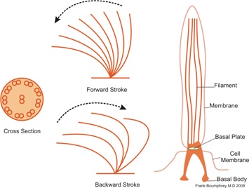
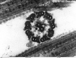
Image Reference: http://commons.wikimedia.org/wiki/File:Cillia1.png
http://commons.wikimedia.org/wiki/File:Chlamydomanas_reinhardtii_Flagella_9_-_TEM.jpg
Types of Locomotion in protozoa
Ciliary movement: It takes place by the presence of cilia that are hair like structure in the animal body. Cilia are shorter and numerous than flagella and are arranged closely in a longitudinal row. They occur throughout the cell surface and beat in co-ordination. This pattern of arrangement is called kinetics. Kinetics denotes the arrangement where complex system is formed between microtubules and the fibres and hence, arises from the basal part called as kinetosomes. In case of ciliary movement, the beating of cilia creates three- dimensional pattern thus working against the viscous force of the water. There are two type of stroke that it performs during ciliary movement: effective stroke and recovery stroke. In effective stroke, the cilia are frequently planar. Each cilium coordinates with the neighboring cilium and this coordination creates a hydrodynamic linkage between the cilia. Thus, a synchronized beat is created and with every beat, layer of surrounding water is displaced. This surrounding water lies parallel to the surface of the cilia. In case of recovery stroke, cilium brush off to the side thus creating a three- dimensional beat. Hence, it sweeping movement is created by the organism that have cilia in an asymmetrical way.
Flagellar movement: Flagella are longer and less numerous that cilia (present at one end) and arise from the basal portion of the protozoan body. In case of flagella; the production of wave along the flagellum produces the force of water that act along the long axis of the flagellum along the direction of the wave. Hence, each flagellum beat independently of the other creating an undulatory movement in symmetrical way.
Amoeboid movement: In case of amoeboid movement, pseudopodia are involved rather than cilia or flagella. In this case, two cytoskeletal proteins called actin and myosin gets polymerized. This creates vacancy and hence cytoplasmic material flow to cover the vacancy that is created due to the polymerization reaction. When amoeba moves, cytoplasm moves to the arm like extension called pseudopodium. This pseudopodium than extends and enlarge and hence this propels the animal body towards that respective direction. Likewise, the movement continues with generation of new pseudopodia and the withdrawal of old pseudopodia takes place. Beside this, the contraction of the posterior side of protozoan body drives the endoplasmic portion forward thus extending its pseudopodium.