Human Skeletal System Assignment Help
We already know that skeletal system means the supporting system of our body. Different systems of our body have particular functions, likewise skeletal system provides framework to our body and helps us in performing different task such as movement, protection and storage. Skeletal system comprises of bones and cartilage. These components functions in a well coordinated manner so as to give shape to our body and aid in its movement.
Structure and physiology
Structure of bone:
Since bone is the main component of the skeletal system, it is important to study depth on its structure. When a bone is examined under the microscope, different parts can be clearly identified.
The Diaphysis: this is the body of any bone i.e. the middle portion, long and cylindrical.
The epiphysis: these are the both ends of the bone, just like two thick, curved ends of any stick.
The metaphysis: this is the main part that helps in growth of the bone. It is located in between Diaphysis and epiphysis. At the growing stage it consist of hyaline cartilage, that helps Diaphysis grow in length, but once the growing age has been crossed, hyaline cartilage is replaced by the bone.
The articulate cartilage: articulation means to join; similarly, articulate cartilage is a layer of hyaline cartilage that joins two bones.
The periosteum: it is present in all those areas where bones are not surrounded by articulate cartilage, thus serving in more than one functions. It helps to protect the bone, nourish bone tissue and repair any fracture if occurred in the bone. It has outer fibrous layer of dense irregular connective tissue and inner osteogenic layer that have cells. Not all, but some of the cells of periosteum help to increase thickness of bone but not its length.
The medullary cavity: it lies within the Diaphysis that has fatty yellow bone marrow.
The endosteum: it is a thin membrane that consists of small layer of cell and connective tissues.
Beside the above mentioned parts, bones also have two different types of tissues: compact and spongy bone tissue that makes the bone.
Compact bone tissue: compact bone also called cortical bone, is one of the strongest bone tissue that makes up the Diaphysis part of the bone. It provides protection, support and resistance against stress that is caused in the bone. This bone tissue has a basic unit called osteon also called Haversian system. Now, each of this Haversian system is made of different parts such as: lacunae, lamellae and canaliculi.
Lamellae: it is the concentric ring made of calcified extracellular matrix, such as mineral salt and collagen fiber. Mineral salt provides hardness whereas collagen fiber provides strength to the bone.
Lacunae: these are the small spaces between lamellae that have osteocytes.
Canaliculi: this are the networks or channels that links lacunaes and hence helps in the transport of minerals to osteocytes and also in removing of any unwanted substances.
Spongy bone tissue: these bone tissue lack osteon and consist of lamellae arranged in irregular lattice of trabeculae. The space between trabeculae is filled with red bone marrow. Trabeculae have lacunae, osteocytes and canaliculi. This type of bone is always covered by compact bone for protection. This bone tissue is light and the trabeculae protect and support the red bone marrow.
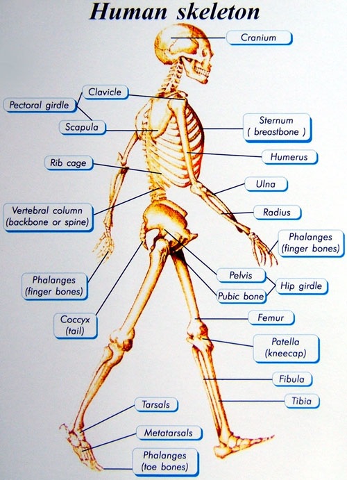
Skeletal system is comprised of 206 bones and all these bones comes under two main parts, axial and appendicular skeleton.

Image Reference: www.endoszkop.com
Axial skeleton: the axial skeleton consists of skull, ribs, sternum, vertebrae and hyoid.
Human Skeletal System Assignment Help By Online Tutoring and Guided Sessions from AssignmentHelp.Net
Skull: skull has mainly two types of bone cranial and facial bone.
Cranial bones are further divided into following types:
1. Frontal bone: this bone is present in the anterior portion of brain. it is made of squama frontalis and Para orbitalis.
2. Parietal bone: it is the large bone that makes top and side of cranium. The external surface of this bone is convex whereas the internal surface in concave with no prominent markings.
3. Temporal bone: it is present at the base and side of the brain
4. Occipital bone: it is the large bone that form base and back of the skull. It has foramen magnum at the inferior part.
5. Sphenoid bone: it is the butterfly shaped bone present in between the frontal and temporal bone.
6. Ethmoid bone: it is a spongy bone present at the top of nasal cavity and in between the orbits.
Facial bones are also divided into different types:
1. Zygomatic bone: these are also called cheekbone that are present forms cheeks and lateral wall of the orbit.
2. Mandible bone: it is present in the lower jaw.
3. Lacrimal bone: it is a small facial bone that forms the portion of anterior medial wall of the orbit.
Vertebrae: these are again subdivided into different types such as: Cervical vertebrae, thoracic vertebrae, lumbar vertebrae, sacrum and coccyx.
1. Cervical vertebrae: atlas is the first bone of cervical vertebrae that supports the skull. Atlas is divided into different parts named anterior arch, anterior tubercle, vertebral foramen, transverse process, posterior arch, posterior tubercule, transverse foramen and superior articulate facet. Likewise, after atlas, axis is the second bone of cervical vertebrae that also have different parts, facet, dens, vertebral foramen, spinous process, lamina and transverse process and foramen. Other bones of cervical vertebrae from 3rd to 9th are almost similar having body, anterior tubercle, posterior tubercle, superior articular process, spinous process, vertebral foramen, transverse foramen.
2. Thoracic vertebrae: 12 thoracic vertebrae are present after the cervical vertebrae in middle of vertebral column. Each of these vertebrae articulate with ribs and have parts that can be well distinguished as: body, superior articular process, vertebral foramen, spinous process, lamina, costal facet and transverse process. All the vertebrae share these common features thus, assisting in formation of a complete vertebral column.
3. Lumbar vertebrae: these are five in number located below the thoracic vertebrae. They are strong and hard. Pedicle, body, vertebral foramen, transverse process, inferior articular process, lamina, spinous process and superior articular process are the parts of lumbar vertebrae.
4. Sacrum: it is a triangular shaped bone that forms posterior portion of pelvis. It is composed of 5 vertebrae. Below sacrum is the coccyx, also called tail bone that has 5-6 small bone s fused together serving in one or different ways.
Ribs and sternum: ribs are twelve in pairs and they articulate with the thoracic vertebrae. Ribs can be divided into different types:
1. True ribs: first seven ribs are called true ribs because they are attached to the sternum directly by costal cartilage.
2. False ribs: 8th to 10th rib that is indirectly attached to the sternum by attachment to the coastal cartilage of rib 7 is called false ribs.
3. Floating ribs: 11th and 12th ribs are called floating ribs because they have no attachment to the sternum.
Sternum: it is also called breast bone that has three segments: manubrium, xiphoid and body. It covers and protects the heart.
Appendicular skeleton: it consist of rest 126 bones of the body. Bones that are mostly from hands and legs constitute appendicular skeleton such as Clavicle, scapula, hummers, radius and ulna, wrist and hand. Pectoral girdle( it is a set of bones that connect upper limb to axial skeleton) consist of 4 bones, 2 clavicle and 2 scapula. Arm and forearms have 6 bones among them 2 are humerus, 2 ulna and 2 radius. Likewise hands have 54 bones that includes 16 carpals, 10 metacarpals, 10 proximal phalanges, 8 intermediate phalanges and 10 distal phalanges. Pelvis has 2 bones, thigh and legs have 8 bones: femur 2, patella 2, tibia 2 and fibula 2. Feet and ankle have 52 bones., 14 tarsals, 10 metatarsals, 10 proximal phalanges, 8 intermediate phalanges and 10 distal phalanges.
Therefore, bones of axial and appendicular altogether makes up the complete skeletal system of human body thus, helping in its movement, protection, support, storage, etc.
Skeletal system is one of the important systems of human body that consist of different bones and cartilages that when work in collaborate manner can help accomplish several tasks effectively and easily. Although individual bones are rigid, the skeleton whole is remarkably flexible. The skeletal serves as an anchorage for the skeletal muscle and as a protective cage for the bodys internal organ. Female bone are usually smaller and lighter than the male bones, similarly, female pelvis is shallower having wider cavity than the male.Therefore, it is important to properly use our body parts and avoid any kind of stress that can result in serious damages.


