HLTAAP001 Recognise healthy body systems Learner Guide
Unit of Competency
Application
This unit describes the skills and knowledge required to work with basic information about the human body and to recognise and promote ways to maintain healthy functioning of the body.
This unit applies to any worker who needs to use and interpret information that includes references to client anatomy and physiology.
The skills in this unit must be applied in accordance with Commonwealth and State/Territory legislation, Australian/New Zealand standards and industry codes of practice.
Unit Mapping Information
No equivalent unit
Performance Criteria
|
Element Elements describe the essential outcomes. |
Performance Criteria Performance criteria describe the performance needed to demonstrate achievement of the element. |
|
1. Work with information about the human body |
1.1 Correctly use and interpret health terminology that describes the normal structure, function and location of the major body systems 1.2 Correctly use and interpret information that relates to the interrelationships between major components of each body system and other structures |
|
2. Recognise and promote ways to support healthy functioning of the body |
2.1 Review factors that contribute to maintenance of a healthy body 2.2 Evaluate how the relationships between different body systems affect and support healthy functioning 2.3 Enhance quality of work activities by using and sharing information about healthy functioning of the body |
Foundation Skills
The Foundation Skills describe those required skills (language, literacy, numeracy and employment skills) that are essential to performance.
Foundation skills essential to performance are explicit in the performance criteria of this unit of competency.
Assessment Requirements
Performance Evidence
The candidate must show evidence of the ability to complete tasks outlined in elements and performance criteria of this unit, manage tasks and manage contingencies in the context of the job role.
There must be evidence that the candidate has:
- Worked effectively with information about the human body and its healthy functioning in at least 3 different situations.
Knowledge Evidence
The candidate must be able to demonstrate essential knowledge required to effectively complete tasks outlined in elements and performance criteria of this unit, manage tasks and manage contingencies in the context of the work role. This includes knowledge of:
- Basic structure and functions of the body systems and associated components, including:
- cells, tissues and organs
- cardiovascular system
- respiratory system
- musculo-skeletal system
- endocrine system
- digestive system
- urinary system
- reproductive system
- integumentary system
- lymphatic system
- nervous system, including sensory systems – eye and ear
- the special senses – smell, taste, vision, equilibrium and hearing
- immune system
- Processes, conditions and resources required by the body to support healthy functioning
- body regulation including:
- maintenance of body temperature
- fluid and electrolyte (including PH) balance
- elimination of wastes from the body
- maintenance of blood pressure
- protection from infection
- physical activity – active and passive.
Assessment Conditions
Skills must have been demonstrated in the workplace or in a simulated environment that reflects workplace conditions. The following conditions must be met for this unit:
- Use of suitable facilities, equipment and resources, including client health information
- Modelling of industry operating conditions, including integration of problem solving
Assessors must satisfy the Standards for Registered Training Organisations (RTOs) 2015/AQTF mandatory competency requirements for assessors
Links
Companion volumes from the CS&HISC website - http://www.cshisc.com.au
Companion Volume implementation guides are found in VETNet - https://vetnet.education.gov.au/Pages/TrainingDocs.aspx?q=ced1390f-48d9-4ab0-bd50-b015e5485705
1. Work with information about the human body
1.1. Correctly use and interpret health terminology that describes the normal structure, function and location of the major body systems
1.2. Correctly use and interpret information that relates to the interrelationships between major components of each body system and other structures
1.1 – Correctly use and interpret health terminology that describes the normal structure, function and location of the major body systems
1.2 – Correctly use and interpret information that relates to the interrelationships between major components of each body system and other structures
By the end of this chapter, the learner should:
- Create a poster detailing the location, structure and function of a range of major body systems.
When working with people in a medical or care context, it is essential to have a good basic knowledge of the body's systems – this will help you understand conditions and recognise concerning symptoms more easily.
Cardiovascular system
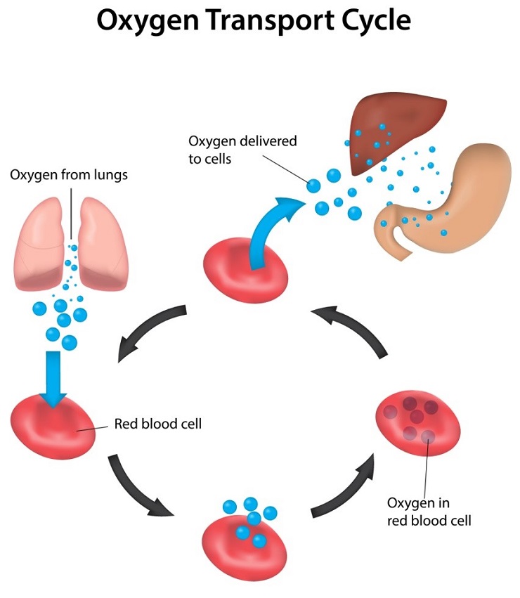
This system consists of the heart, blood vessels and around five litres of blood that it transports around the body. It is responsible for transporting oxygen, hormones, nutrients and waste products around the body. The heart powers this system, pumping the blood which carries the previously mentioned contents at a rate of five litres per minute.
The heart
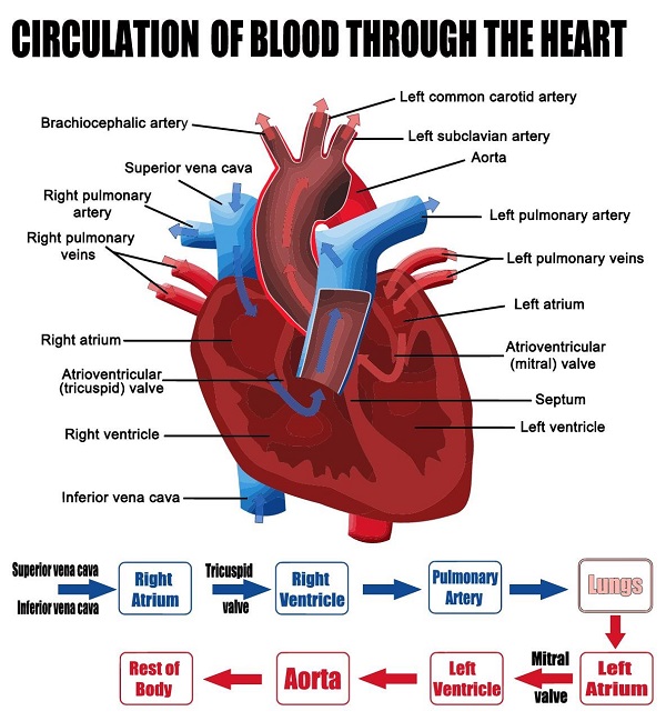
This is a muscle that is located in the thoracic region, between the lungs. Two-thirds of the heart is on the left side of the body; the top of the heart is connected to the aorta, vena cava, pulmonary trunk and pulmonary veins – the major blood vessels. It is a four-chambered 'double pump' where the left and right sides function separately – the right side pumps deoxygenated blood and the left side oxygenated blood. Each heartbeat pumps both sides of the heart simultaneously.
Circulatory loops
In the human body, there are two circulatory loops:
- Pulmonary circulation loop – transports deoxygenated blood from the right side of the heart to the lungs; here, the blood becomes re-oxygenated and is transported back to the left side of the heart. The right atrium and right ventricle pump blood along this loop.
- Systemic circulation loop – transports oxygenated blood from the left side of the heart to all body tissue (apart from the heart and lungs). It also removes waste from tissue and returns deoxygenated blood to the right side of the heart. The left atrium and left ventricle pump blood along this loop.
Blood vessels
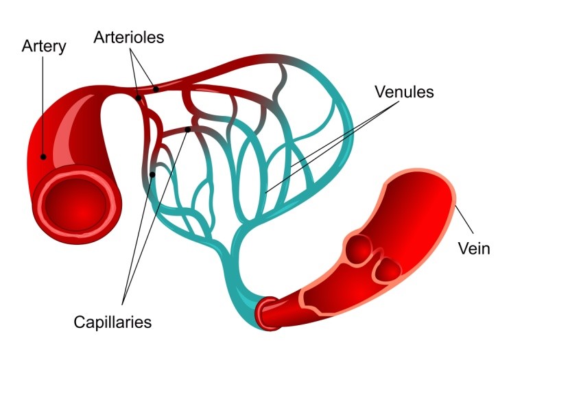
These allow blood to travel from the heart to every area of body and back – they are sized according to how much blood passes through that particular area of the body. Blood travels through a hollow area called the lumen, which is encased in a wall (thin for capillaries and thick for arteries). The blood vessels are lined with endothelium which keeps blood inside of them and prevents the formation of clots
There are three types of blood vessels:
- Arteries – they carry blood away from the heart, which is usually oxygenated; the only exception is the pulmonary trunk circulation loop, which carries deoxygenated blood from the heart to the lungs. They have high levels of blood pressure, as the blood is being pushed from the heart (hence the thicker vessel walls). Bigger arteries are more elastic, to accommodate the pressure, whereas smaller arteries are muscular and contract/expand to regulate blood flow.
Arterioles are narrow arteries that branch off from the ends of arteries and carry blood to capillaries. They have lower blood pressure as they are greater in number, further from the heart and carry less blood per unit – therefore, the walls are much thinner than arteries. They also use muscle to regulate blood flow.
- Capillaries – these are the smallest, thinnest and most common blood vessels in the body. They run through just about every tissue in the body and connect arteries to venules. In capillaries, gases, nutrients and waste products are exchanged between tissues – therefore, the endothelium is thin to allow easier passage of materials, while still retaining blood cells inside the vessels.
Precapillary sphincters regulate blood flow into capillaries by reducing blood flow to inactive tissue and directing it towards active tissue.
- Veins – they return blood to the heart; their walls are much thinner, less elastic and less muscular than arteries (as they are not subjected to as much pressure). Instead, gravity, inertia and skeletal muscle contractions allow veins to push blood back to the heart. Many veins contain valves that prevent blood from going away from the heart.
Venules perform the same function as arterioles but collect blood from capillaries, rather than deposit it.
Functions of the cardiovascular system
- Transportation of blood (along with its contents) to body tissue
- Protection – white blood cells clean up debris and fight infections; platelets and red blood cells form scabs that prevent infection and open wounds; blood also carries antibodies to help protect from specific infections
- Regulation – helps maintain body temperature; helps balance the body's pH; maintains concentration of body's cells.
Regulation of blood pressure
- The greater the contractions of the heart, the higher the blood pressure
- Vasoconstriction – the diameter of an artery is reduced by contracting the muscle in the arterial wall. Blood pressure is increased and flow reduced.
- Vasodilation – the diameter of an artery is increased as the muscle in the arterial wall relaxes. This can be caused by hormones or chemicals, artificial or natural.
- An increase in blood volumes equals higher blood pressure
- Thicker blood (from clots etc.) raises blood pressure.
Normal blood pressure is recommended at 120 over 80mmHg; this level can help to lower the risk of having heart disease or a stroke (sourced from, ‘What is normal blood pressure?’ at Blood Pressure UK: http://www.bloodpressureuk.org/BloodPressureandyou/Thebasics/Whatisnormal access date: 20.03.2017).
A healthy blood pressure ensures that blood is pumped around the body efficiently, providing energy and oxygen throughout.
Haemostasis
This is where blood clots and forms scabs – it is controlled by the platelets of the blood. Platelets remain inactive until they reach damaged tissue and they change form to a spiky shape and become sticky in order to hang onto damaged tissue. They then release chemicals to produce fibrin, which forms the structure for a blood clot. They will stick together to plug a wound until it can be fully repaired, protecting it from foreign bodies in the meantime.
Respiratory system
This system provides oxygen to the cells of the body and removes carbon dioxide from them.
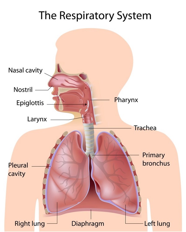
It consists of three major areas:
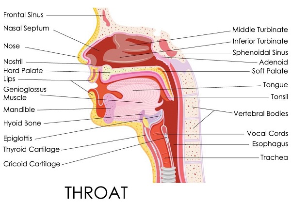
- The airway – nose, mouth, pharynx, larynx, trachea, bronchi, and bronchioles
- The lungs – pass oxygen into the body and carbon dioxide out of it
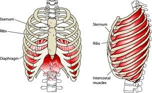
- Respiration muscles – diaphragm, intercostal muscles.
Nose/nasal cavity
This nasal cavity is the primary tract through which air moves; the nose is made of cartilage, bone, muscle and skin, and protects the nasal cavity. The nasal cavity warms, moisturises and filters air that enters the body before it goes to the lungs. Hairs and mucus trap dust and other contaminants. Exhaled air returns moisture and heat to the nasal cavity before exiting the body.
Mouth
This is the secondary tract through which breathing takes place and is used when extra air is needed. However, it doesn't warm and moisturise air as well as the nose and doesn't filter as well. However, it allows more air to enter the body quicker.
Pharynx
This is the throat and is a muscular funnel that goes from the end of the nasal cavity to the oesophagus and larynx. It contains the epiglottis, which is a flap of cartilage that moves between the trachea and oesophagus, blocking the correct passage, depending if you are eating or not – this prevents choking.
Larynx
This is the voice box and contains vocal cords, the epiglottis and is constructed of cartilage.
Trachea
This is the windpipe and is made of cartilage rings – it connects the larynx to the bronchi and allows passage of air into the lungs – it contains mucus to trap external bodies from reaching the lungs. This mucus is then moved toward the pharynx, where it is swallowed and digested.
Bronchi/bronchioles
This is where the airway splits into two branches, which then spilt into secondary branches (two in the left lung, three in the right lung). Secondary bronchi then spilt into tertiary bronchi and then into bronchioles, which further split until they become less than a millimetre in diameter – these are known as terminal bronchioles and transfer air into the alveoli of the lungs.
Muscle tissue in the bronchi and bronchioles helps regulate airflow – they relax when more air is required (e.g. during exercise) and contract when resting to prevent hyperventilation.
Lungs
The lungs are organs and are surrounded by a pleural membrane to allow expansion and a negative pressure space to allow for passive filling of the lungs as they relax. The left lung is slightly smaller, to accommodate the heart and only has two lobes, comparative to the right lung's three.
They contain around 30 million alveoli, which are tiny cup-shaped structures that allow the exchange of gases between the air in the lungs and the blood passing through the capillaries.
Muscles of respiration
There are muscles surrounding the lungs that allow air to be inhaled and exhaled from the lungs. The primary muscle responsible for this is the diaphragm – situated at the floor of the thorax. When it contracts, it moves into the abdominal cavity and allows air to be pulled into the lungs; relaxation of the muscle allows air to flow back out of the lungs. Intercostal muscles between the ribs assist the diaphragm in expanding and compressing the lungs.
Types of respiration
There are two types of respiration:
- External respiration – the exchange of gases from the air into the blood
- Internal respiration – the exchange of gases between blood and the tissues of the body.
Homeostatic control of respiration
When the body is resting, it maintains what is called eupnoea – this is a steady breathing rate and happens during rest. When we become active, the body requires more oxygen and is producing more carbon dioxide – therefore, chemoreceptors send signals to the brain which will instruct the body increase its rate and depth of breathing to cope with the situation.
Musculo-skeletal system
(Source: Understanding the basic anatomy and physiology of the ... (n.d.). Retrieved from http://lrrpublic.cli.det.nsw.edu.au/lrrSecure/Sites/LRRView/7700/documents/5657/)
This is the structure of the body and consists of the bones, muscles, joints and the tendons and ligaments that hold them all together.
The function of it is to:
- Protect and support the organs of the body and its internal structures
- Allow movement
- Give shape and structure to the body
- Produce blood cells
- Store calcium and phosphorus
- Produce heat.
The skeletal system
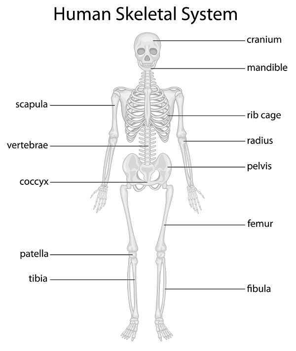
There are 206 bones in the human body – the skeletal system of bones and joints as the supporting structure of the body.
Bones are made from calcium-phosphorus, organic matter and water. They are covered in periosteum, a living membrane that houses osteoblasts, which contain bone forming cells. The centre of bone contains marrow, which consists of fat cells, blood vessels and blood cell manufacturing tissue.
Bones can be in one of four shapes:
- Flatg. ribs
- Irregularg. vertebrae
- Shortg. carpals
- Longg. humerus.
Joints allow movement and are where two or more bones are held together by ligaments.
There are three types:
- Fibrous (immovable)g. skull
- Cartilaginous (slightly moveable)g. vertebrae
- Synovial (freely moveable):
- ball and socketg. hip
- hingeg. knee
- glidingg. wrist (carpals)
- pivotg. radius, ulna
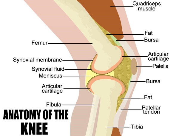
There are various types of movement that the bones of these joints can perform:
- Abduction – away from the body
- Adduction – towards the body
- Flexion – bending a limb towards the body
- Extension – extension of a limb away from the body
- Rotation – movement around a point.
The muscular system
Muscles provide the contractions and relaxations that move the bones around the joints, working in conjunction with the skeletal system. There are over 500 muscles in the body; they also help maintain the position of the body.
Tendons attach muscle to bone; there are three types of muscles:
- Skeletal – also known as voluntary, these attach muscles to bones
- Smooth – also known as involuntary, they control internal processes such as the action of the gut and blood vessels
- Cardiac – in the heart.
The contraction of a muscle shortens it – this is caused by the release of chemicals by the brain and the trigger of a motor nerve. The contraction pulls the bone with it; as this happens, the antagonist muscle relaxes to facilitate the movement – these are known as antagonistic pairs.
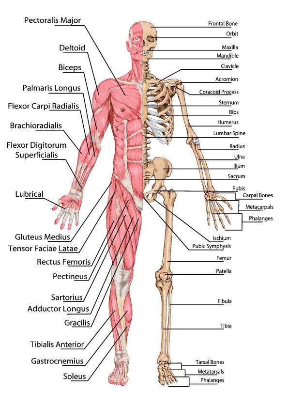
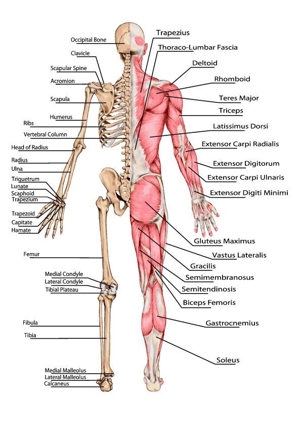
The following two diagrams detail all the major bones and muscles in the body:
Endocrine system
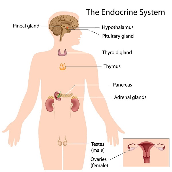
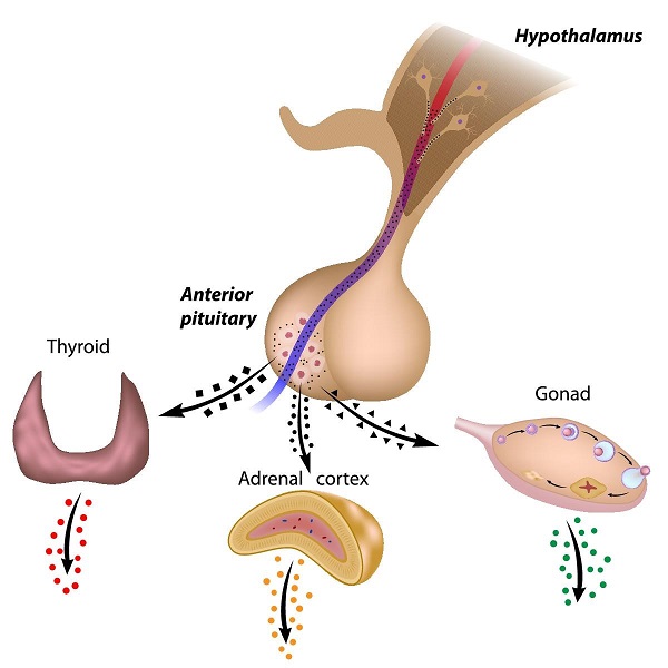
This system consists of all the glands in the body and the hormones they produce. The glands are controlled by the nervous system and chemical receptors in the blood. They help maintain homeostasis (stable internal condition) by regulation of organ functions. Hormones are responsible for things like metabolism, sexual development and reproductions, mineral and sugar retention, heart rate and digestion.
Hypothalamus
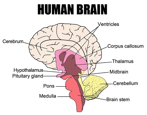
This part of the brain directly controls the endocrine system through the pituitary gland; it is also responsible for various nervous system-related jobs. It contains neurosecretory cells – these are neurons that secrete releasing and inhibiting hormones. These hormones are responsible for the controlled release of things like growth hormone and follicle stimulating hormone.
Pituitary gland
This is a pea-sized piece of tissue connected to the hypothalamus, which releases hormones through blood vessels surrounding it.
It is made of two parts:
- Posterior pituitary – releases oxytocin (for childbirth contractions and release of milk for breastfeeding) and antidiuretic hormone (prevents water loss in body by reducing blood flow to sweat glands and increasing water uptake in kidneys)
- Anterior pituitary – controlled by the hypothalamus, it produces six vital hormones:
- thyroid stimulating hormone (TSH)
- adrenocorticotropic hormone (ACTH)
- follicle stimulating hormone (FSH)
- luteinising hormone (LH)
- human growth hormone (HGH)
- prolactin (PRL).
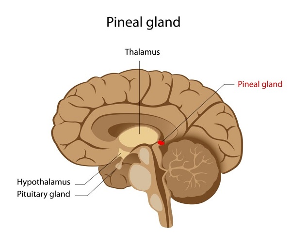
Pineal gland
This produces melatonin, to help regulate the sleep-wake cycle – increased production causes feelings of drowsiness.
Thyroid gland
Located at the base of the neck around the lateral sides of the trachea, it produces:
- Calcitonin – regulating blood calcium levels
- Triiodothyroxine and thyroxine – regulating metabolic rate.
Parathyroid glands
They produce parathyroid hormone (PTH) when calcium ion level drop too low – this stimulates the osteoclasts to break down the calcium stores from bones, so they are released into the bloodstream. It also triggers kidneys to return calcium back into the bloodstream.
Adrenal glands
Found above the kidneys, they are made of two layers:
- Adrenal cortex
- produces glucocortoicords (breaks down proteins and lipids; reduces inflammation and triggers immune response)
- mineralocorticoids (help regulate mineral ions concentration in the body)
- androgensg. testosterone (regulate growth and activity of cells)
- Adrenal medulla – produces epinephrine and norepinephrine, helping increase blood flow to the brain and muscles under stress (known as 'fight or flight' reaction). They increase heart rate, breathing rate and blood pressure, while decreasing blood flow to organs that are not involved in responding to emergencies.
Pancreas
This is a large gland near the stomach which releases glucagon to raise blood glucose levels and insulin to lower them after eating.
Gonads
These are the ovaries (in females) and testes (in males), which produces sex hormones – they determine the respective secondary sex characteristics of adults.
- Testes – releases testosterone (causes growth and increases in strength of the bones and muscles, particularly during puberty; causes inherited hair loss; triggers sexual development)
- Ovaries – release oestrogen and progesterone (for ovulation and pregnancy; for sexual and growth development during puberty)
Thymus
Found behind the sternum, it produces thymosins (to develop t-lymphocytes during foetal and child development). During puberty, it becomes inactive and is replaced by adipose tissue.
Other hormone producing organs
- Heart – atrial natriuretic peptide (ANP) in response to high blood pressure levels
- Kidneys – erythropoietin (EPO) in response to low levels of oxygen in the blood
- Digestive system – cholecystokinin (CCK), secretin, and gastrin are all produced by the organs of the gastrointestinal tract
- Adipose – produces the hormone leptin that is involved in the management of appetite and energy usage by the body
- Placenta – human chorionic gonadotropin (HCG) assists progesterone by signalling the ovaries to maintain the production of oestrogen and progesterone throughout pregnancy
- Local hormones – prostaglandins and leukotrienes are produced by every tissue in the body (except for blood tissue) in response to damage.
Nervous system
This is akin to the electrical wiring system of the body and is comprised of nerves that go from the brain to every part of the body.
Neurons send signals through thin fibres which cause chemicals (neurotransmitters) to be released at junctions known as synapses. These give a command to a cell to behave in a certain way – this whole process takes about a fraction of a millisecond.
Sensory neurons react to stimuli such as light, sound and touch – they then send feedback to the brain through the central nervous system, communicating about the surrounding environment. Motor neurons transmit messages to activate muscles/glands.
Neurons are help in place by glial cells (neuroglia), which also destroy pathogens, remove dead neurons and ensure the signals sent by the brain reach their intended target.
The brain
This soft organ is located inside and protected by the skull – it is the main control centre of the body and contains 100 billion neurons. Along with the spinal cord, it is part of the central nervous system – it is responsible for things like consciousness, memory, decision-making, involuntary and voluntary contractions.
The spinal cord
This is a long, thin column of neurons bundled together – it carries information down and around the body, resulting in conscious movement, as well as reflexes.
Nerves
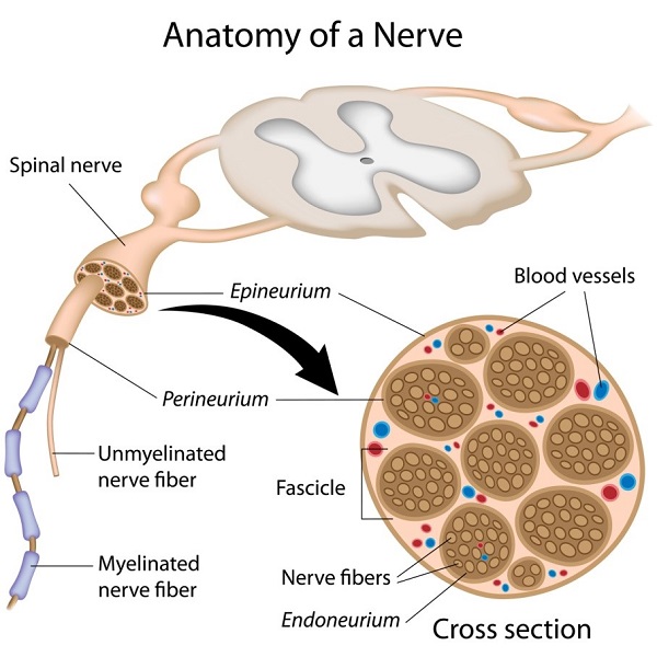
Nerves are bundles of axons that are information highways – these bundles are known as fascicles and are wrapped in a protective layer called the perineurium; groups of these fascicles are wrapped together to form an entire nerve.
Digestive system
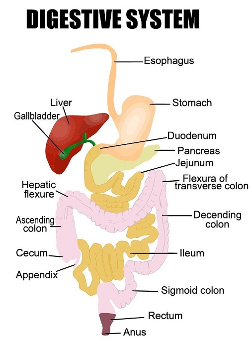
This is made up of the gastrointestinal (GI) tract, liver, pancreas and gallbladder. In the GI tract, tubes join from the mouth to the anus, so that food and drink which is ingested is digested and leaves the body as faeces, once all useful nutrients have been extracted for use in the body.
These useful nutrients include:
- Carbohydrates – they can be simple and complex; they are used for energy and make up 45-65 percent of the total daily calorie intake.
- Protein – they are broken down into amino acids, which are the building blocks of the body and are used for growth and repair.
- Fats – these are a rich source of energy and help absorb vitamins in the body. They make up 20-35 percent of daily calories; they are broken down into fatty acids and glycerol. The body also stores excess energy as fat.
- Vitamins – they can be water-soluble and fat-soluble; each vitamin has a different role in the growth and health of the body. Fat-soluble vitamins are stored in fat, whereas water soluble vitamins are flushed out in urine.
There are bacteria in the GI tract called gut flora that help the digestion process, along with the nervous and circulatory systems.
Food moves through the GI tract via peristalsis (the movement of organ walls), which also allows the contents to be absorbed.
The organs involved in the digestive system include:
- Mouth – for chewing and breaking down starches
- Oesophagus – for swallowing
- Stomach – for letting food enter and mix it with digestive juices; it breaks down protein
- Small intestine – for peristalsis and breaking down starches, protein and carbohydrates
- Pancreas – to break down starches, fats and protein
- Liver – to break down fats
- Large intestine – to change waste products into faeces and to excrete it from the body during a bowel movement.
Digestive juices include:
- Saliva – produced by salivary glands. It contains enzymes and moistens food to allow it to move more easily through the oesophagus
- Stomach enzymes – produced by the glands in the stomach lining, they digest protein
- Pancreas enzymes – these break down carbohydrates, fats and proteins
- Bile – produced by the liver and stored in the gallbladder, it dissolves fat, so it becomes digestible
- Small intestine enzymes – they combine with pancreatic juice and bile to complete the digestive process, breaking down proteins and starches to make glucose molecules.
Urinary system
This is the body's system for removing urine (made up of waste and excess fluid). For urination to occur, it requires all of the required parts of the urinary tract to work in sequence.
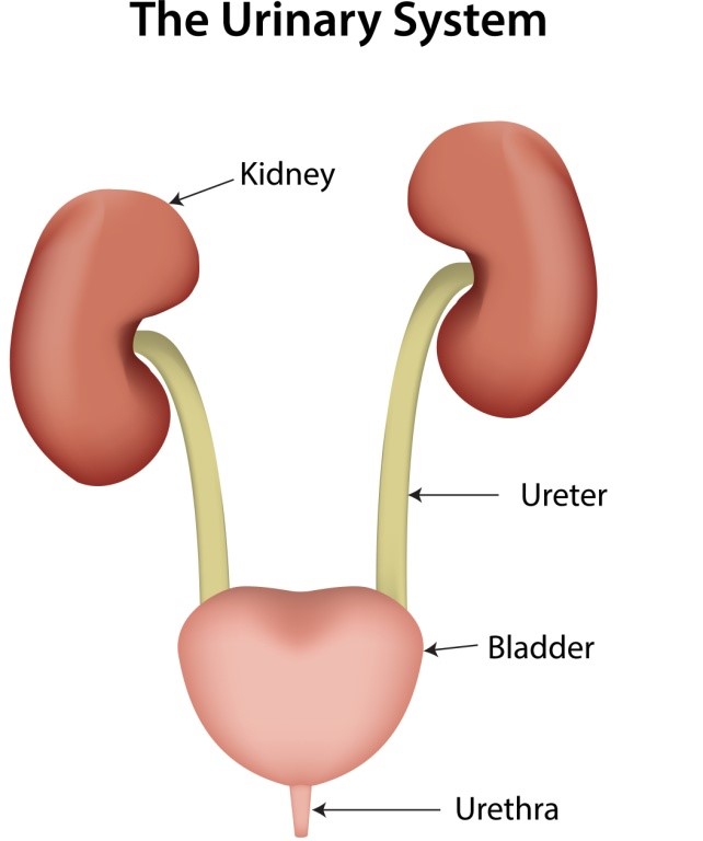
The parts of the urinary tract include:
- Kidneys – they filter about 240 to 300 pints of blood to make two to four pints of urine
- Ureters – they carry urine from the kidneys to the bladder
- Bladder – it expands as it fills with urine, and the emptying of it is controlled by the person. A normal bladder can hold around one and a half to two cups of urine; when it fills to capacity, signals are sent to the brain to tell them to find a toilet. During urination, the bladder empties through the urethra. The sphincters (internal and external) control whether urine stays in the bladder or exits through the urethra.
The kidneys function to:
- Prevent the build-up of waste and extra fluid in the body
- Keep electrolyte levels stable
- Make hormones to regulate blood pressure
- Make red blood cells
- Keep bones strong.
Reproductive system
These are a collection of organs that combine with the purpose of creating new life (making babies). The reproductive organs include genitalia and internal organs, such as the gonads.
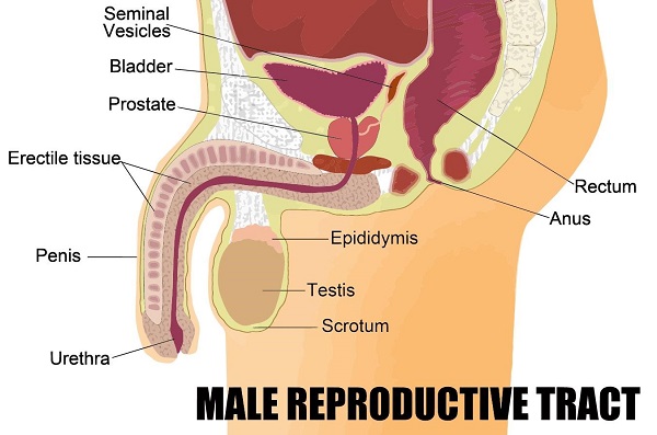
The male reproductive system consists of:
- Testes – encase in the scrotum, they contain sperm, which are the male sex cells. They also make male sex hormones, for sexual development
- Glands – they create fluids that mix with sperm
- Sperm ducts – the sperm travel through these and are mixed with fluids from the glands, created semen
- Urethra and the penis – the urethra can either pass out urine or semen of the body, depending on whether you are urinating or having sexual interAssignment.

The female reproductive system consists of:
- Ovaries – they contain hundreds of unfertilised eggs (ova). A woman has these cells from birth (compared to the continual production of sperm in men).
- Fallopian tubes – these connect the ovaries to the uterus. Every month, when an ovary releases an egg, they are transferred through the fallopian tubes to the uterus.
- Uterus – also known as the womb, this muscular organ is where fertilised eggs develop into babies.
- Cervix – this is a ring of muscle on the lower part of the uterus – it serves to hold the baby in during pregnancy.
- Vagina – this is a muscular tube the goes from the cervix to the outside of the body on a woman. This is also the entrance for a man's penis during sexual interAssignment. The opening of the vagina has a vulva (two folds of skin, called labia). The urethra opens into the vulva and is the exit for urine; however, it is a separate entity to the vagina.
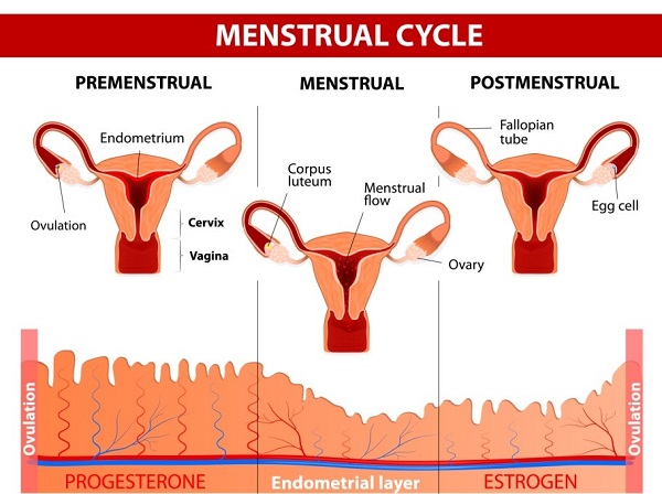
The menstrual cycle
This 28-day cycle begins with bleeding of the vagina – this is the lining of the uterus that has been lost. This is known as having a period or menstruation. This continues for around five days. The uterus lining begins to re-grow and an egg cell begins to mature in an ovary. On day 14, the mature egg cell is released into the fallopian tube, towards the uterus (known as ovulation). If the egg is not fertilised by a sperm cell, the lining of the uterus breaks down and the cycle repeats; if it is fertilised, the egg attaches to the lining of the uterus and the woman becomes pregnant.
Fertilisation
When a man ejaculates into a woman's vagina during sexual interAssignment, the sperm cells travel to the uterus through the cervix; there, if it meets with an egg, fertilisation happens and an embryo forms from the fertilised egg. This then develops into a foetus and then a baby.
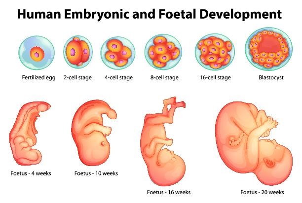
Foetus development
The foetus requires protection, nutrients and oxygen to survive – these are all provided by the mother; it also needs waste products to be removed.
- Protection – this is done by the uterus and amniotic fluid
- Oxygen and nutrients – this is provided by the placenta
- Waste removal – this is also performed by the placenta
The placenta grows in the wall of the uterus and is connected to the baby via an umbilical cord – it lets substances pass between the blood supplies of the mother and baby via diffusion but never lets the blood mix together.
Birth
After nine months, the baby is fully developed and ready to be born and the cervix relaxes as the uterus wall muscles contract to push the baby out of the body.
Puberty
As a child grows into an adult, they go through puberty (between the ages of ten and 15) – where their reproductive system develops so that they can produce children of their own.
Some of the other changes include:
- Growth of underarm hair
- Growth of pubic hair
- Body smells become stronger
- Increased rate of bone and muscle growth
- Emotional development.
In males, the following changes happen:
- The voice breaks
- Testes start producing sperm
- Shoulders get wider
- Testes and penis enlarge
- Hair growth on the face and chest.
In females, the following changes happen:
- Development of breasts
- Ovaries start releasing eggs and periods begin
- Hips get wider.
Integumentary system
This system consists of the skin, the largest organ in the body. It protects the internal parts of the body from damage, prevents dehydration, stores fat and produces hormones and vitamins. By assisting with body temperature and water regulation in the body, it helps maintain homeostasis. It is also the first defence measure against bacteria, viruses and other harmful microbes, as well as ultraviolet radiation.
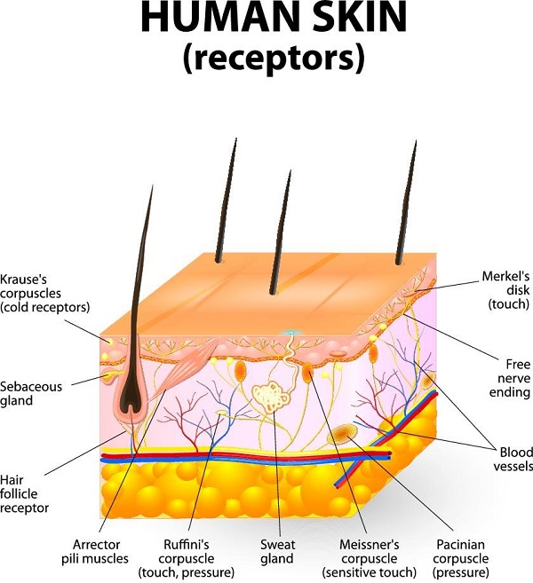
The skin has receptors that detect heat and cold, pain, pressure and touch.
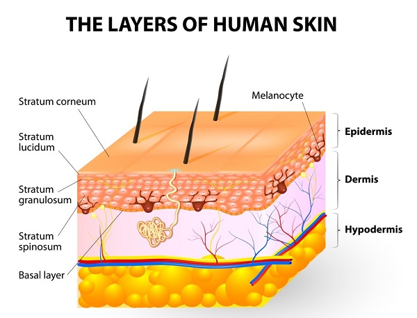
The components of the skin are:
- Hair
- Nails
- Sweat glands
- Oil glands
- Blood vessels
- Lymph vessels
- Nerves
The anatomy of the integumentary system consists of:
- The epidermis – this is comprised of squamous cells which create keratin – this is major component of skin, hair and nails. It is the outermost layer of the skin. It can either be thick (on the palms of hands and feet) or thin (rest of the body) skin.
- Dermis – this is the thickest layer of the skin (90 percent) and it supports the epidermis. It contains special cells that help regulate temperature, fight infection, store water and supply nutrients and blood to the skin. They also detect sensations and give skin its strength and flexibility.
It contains blood vessels, lymph vessels, sweat glands, sebaceous glands, hair follicles, sensory receptors, collagen and elastin.
- Hypodermis – this innermost layer of the skin helps insulate the body and cushions the internal organs. It is composed of fat and loose connective tissue and connects skin to underlying tissues through collagen, elastin and reticular fibres from the dermis. It contains a specialised tissue called adipose that stores excess energy as fat. Blood vessels, lymph vessels, nerves and hair follicles also extend through this layer of the skin.
Modified from: Integumentary System - About.com Education. (n.d.). Retrieved from http://biology.about.com/od/organsystems/ss/integumentary_system.htm
Lymphatic system
This system's primary function is to transport lymph, which is a clear and colourless fluid that contains white blood cells (to fight disease). Lymph helps the body get rid of toxins, waste and other unwanted substances from the body. It also transports fatty acids from the intestines to the circulatory system.
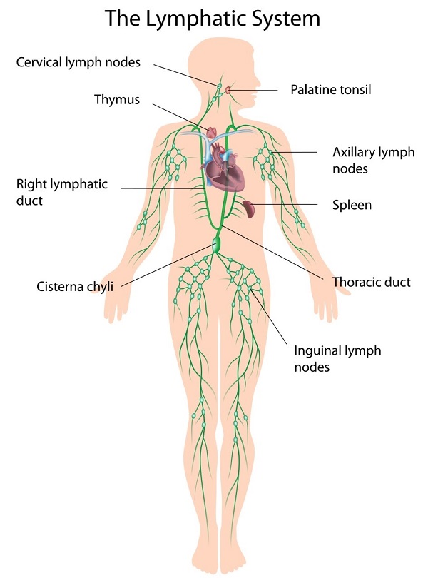
The lymphatic system consists of:
- Lymph vessels – they carry lymph through the body and they resemble veins in structure.
- Lymph nodes (600 to 700) – make more white blood cells, to fight infection.
- Lymph – flows upward towards the neck, through the subclavien It is created from any fluid that doesn't return to the heart via the veins.
- Tonsils – clusters of lymphatic cells in the pharynx. They are commonly removed after persistent throat infections.
- Spleen – helps protect against infection and is just above the kidney. Humans can live without this, but are more prone to injury and infection without it.
- Thymus – in the chest, just above the heart; it stores immature lymphocytes and prepares them to become active T cells.
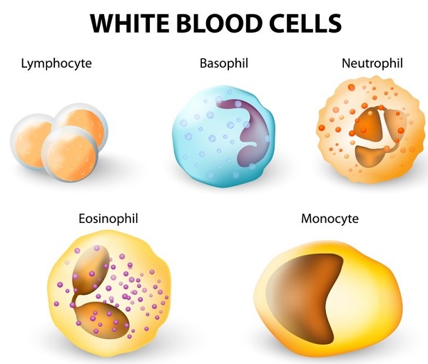
The special senses – vision, hearing, equilibrium, smell and taste
- Vision – initiated by light interacting with sensory receptors in the retina of the eye
- Equilibrium – initiated by motion interacting with sensory receptors in the ear
- Hearing – initiated by sound waves interacting with sensory receptors in the ear
- Smell – initiated by chemicals interacting with sensory receptors in the nose
- Taste – initiated by chemicals interacting with sensory receptors in the tongue.
The eye
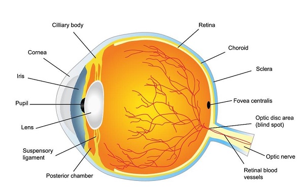
The eye is composed of three layers:
- The sclera – the outermost layer of white, connective tissue – it contains the cornea (a transparent area of the eye that allows light to enter the eye
- The choroid – the second layer, containing blood vessels and pigment cells
- The retina – the innermost layer, containing the light sensitive cells, cones and rods.
Another component of the eye is:
- The iris – the coloured part of the eye that controls how much light enters the eye.
The ear
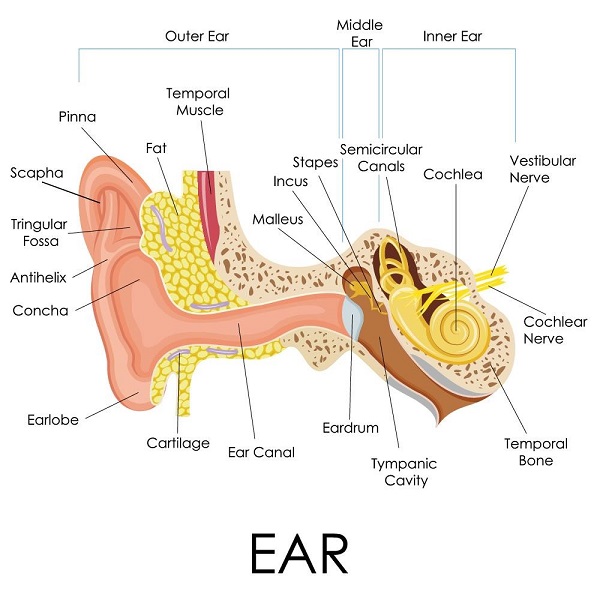
- The external, middle and inner sections of the ear contain the organs for balance and hearing.
- The visible part of the ear is called the auricle – it directs sound waves to the ear canal.
- The ear canal is lined with hairs and ceremonious glands that create earwax to protect the eardrum.
- The middle ear contains bones called ossicles – the hammer, the anvil and the stirrup. They transmit sound vibration to the oval window from the timpanic membrane (the connection with the inner ear).
- The middle ear contains the Eustachian tube, which connects to the pharynx and allows equalisation of air pressure between external and internal environments.
- The inner ear contains fluid and interconnecting chambers/tunnels in the temporal bone. The cochlea is responsible for hearing and the semicircular canals and vestibule for balance.
The nose
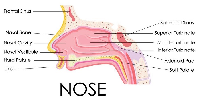
- Molecules of air are dissolved in the mucus lining of the nose; these molecules are then transferred to the olfactory bulb and cortex in the temporal and frontal lobes of the cerebrum. This creates smell.
The mouth
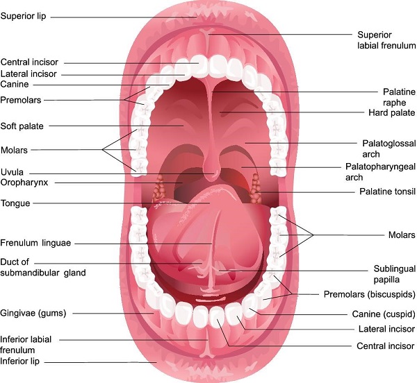
- Taste buds are found on the tongue, the palate of the roof of the mouth and part of the pharynx.
- Taste buds are made up of epithelial cells on the exterior and taste cells in the interior.
- Taste is determined when the chemical is dissolved in saliva and transferred to the taste cortex in the parietal lobe of the brain via the facial, glosspharingeal and vagus nerves.
Cells, tissues and organs
Cells form tissues, tissues form organs and organs combine into organ systems.
Cells
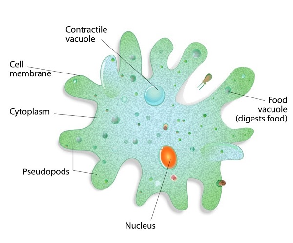
Cells are the building blocks of all organisms (including animals and plants).
An animal cell features a nucleus, cell membrane, cytoplasm and vacuole. However, they do not have a cell wall or chloroplasts like a plant cell.
- The nucleus – contains all the genetic information to make a copy of the cell/organism – it controls the activities of the cell
- The cell membrane – this is a filter that allows certain substances in and out of the cell (e.g. water and oxygen)
- Cytoplasm – it is water-based and where the cell reactions occur
- Vacuole – this contains a watery solution where substances are stored (including waste).
Cells can become specialised to carry out different tasks e.g. the lung lining cells have tiny hairs to move mucus towards the mouth.
Tissues
Tissues are formed by cells with similar structures and functions combining to form a group.
Examples of tissues are:
- Muscle
- The lining of the lungs
- The intestinal lining.
Organs
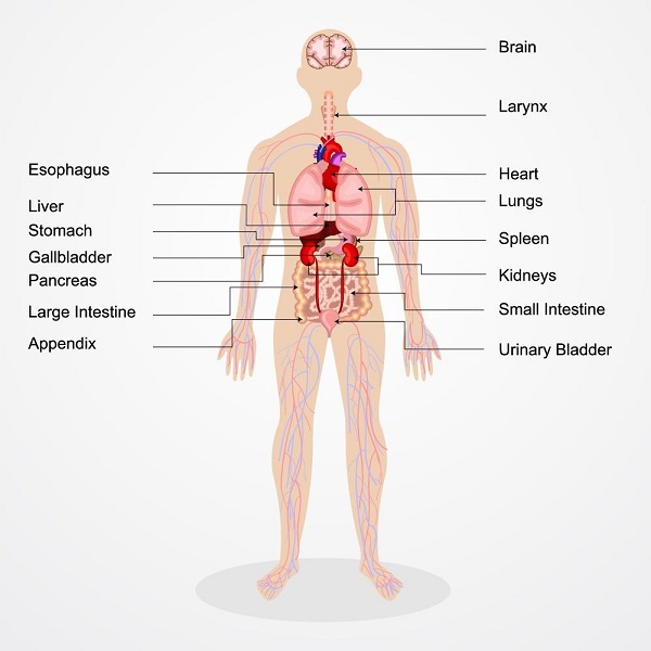
Organs of the human body
Organs are made from groups of different tissues, which work together to do a particular job.
Examples of organs are:
- The heart
- Lungs
- The brain.
Fluid and electrolytes in the body
Fluid is essential to help our body carry out all its functioning. Our bodies require fluid to hydrate, to transport blood cells around the body and to enable cellular activity to take place. Every action our body carries out will be supported and enabled by fluid.
Electrolytes are minerals in our body fluids; they carry an electric charge which keeps the heart, nerves and muscles functioning as they should; they also help to maintain our blood pH levels. The kidneys help to maintain balance with our electrolyte values, when electrolyte values are not balanced this can damage our health.
Common electrolytes in the body are:
- Calcium
- Sodium
- Potassium
- Phosphate
- Chloride
They also help ensure fluid levels inside and outside of cells are in balance; cells can change electrolyte values by drawing more fluid in or eliminating fluid.
Information on electrolytes has been sourced from ‘The Role Of Electrolytes In The Body’ by Sonia Gulati.
The importance of maintaining pH balance
Our body is affected by changes in pH levels; if our pH levels become too acidic, it decreases our oxygen levels in the blood and creates the right environment for disease. We should maintain an alkaline pH balance to maintain healthy functioning; this is recommended as a pH of 7.35. A high acid pH level will encourage the body to use its store of calcium in the bones to reduce the levels to a more alkaline one.
Information on pH balance has been sourced from ‘Maintain proper pH balance’ at Enviro-Health Tech: http://www.envirohealthtech.com/ph.htm (access date: 20.03.2017).
Activity 1A
2. Recognise and promote ways to support healthy functioning of the body
2.1. Review factors that contribute to maintenance of a healthy body
2.2. Evaluate how the relationships between different body systems affect and support healthy functioning
2.3. Enhance quality of work activities by using and sharing information about healthy functioning of the body
2.1 – Review factors that contribute to maintenance of a healthy body
By the end of this chapter, the learner should:
- Create a guide which reviews how to maintain a healthy body
- Create an exercise schedule for a person over 65, considering physical limitations, safety concerns and time restraints.
Maintaining a healthy body
A healthy body is not something that is a given – there need to be basic maintenance strategies to promote health and proper function of all the essential body systems.
Exercise
Regular exercise should be taken to ensure that the musculoskeletal system is maintained. The recommended amount is at least thirty minutes of moderate intensity per day for young healthy people – of Assignment, as people age, this becomes less possible due to movement and joint problems.
Active exercise
This is where there is voluntary movement and force exerted by the muscles of someone – this includes anything from running, to weight lifting, to standing up.
Passive exercise
This is where there is force exerted on muscles involuntarily e.g. range of motion exercises, performed by a nurse or a physio.
Recommended exercise activities (relative to age and physical condition) include:
- Walking
- Jogging
- Interval training
- Weight training
- Aerobics
- Swimming
- Cycling
- Yoga
- Pilates
- Hiking
- Dancing
Mental exercise
As well as maintaining the condition of your bones, muscles and organs, you also need to take care of the most vital organ of all – the brain.
This involves performing mentally stimulating exercises, and can be very effective at delaying the effects or presentation of Alzheimer's disease and dementia (however, it cannot prevent it).
Examples of mentally stimulating activities include:
- Reading
- Doing a puzzle
- Crosswords
- Learning a new skill
- Problem-solving activities
- Writing letters (or other texts)
- Playing chess
- Memory recall activities
- Performing activities using a non-dominant limb
- Activities using multiple senses simultaneously.
Another way of mentally stimulating yourself is to break out of habitual routines. Simply doing daily tasks in a different order, taking different routes, etc. will force you to think about what you are doing, rather than simply living in 'autopilot'.
Medical check-ups
It is recommended that you have regular check-ups from the age of 20 and above – these can include a variety of tests to make sure that the internal organs and musculoskeletal systems are working correctly.
Some of the tests include:
- A complete physical – have one every five years from the ages of 20-45; every two years from 45-65; and every year thereafter.
- Dental check-ups – teeth should be cleaned and checked by a dentist every six months to a year. If you smoke or chew tobacco, these exams are even more important.
- Eye exam – if you wear glasses or contact lenses, you should have an eye exam every two years, or yearly if you have From the age of 40, you need a complete eye exam every two years.
- Colon exam – you should get your colon tested from around the age of 40 upwards, as colon cancer is the third most common form of cancer:
- yearly faecal occult blood test (FOBT)
- a flexible sigmoidoscopy every five years
- double-contrast barium enema every five years
- colonoscopy every 10 years.
- Prostate check (for men) – for men over 60, or 50 if a family history of prostate cancer exists.
- Skin check – for melanoma and non-melanoma (every two to three years from the age of 20 and every year after the age of 40.
- Blood pressure check – every two years (or every year if you have high blood pressure).
- Electrocardiogram (to test for heart disease) – get one at age 40.
- Cholesterol check – every five years.
- C-Reactive Protein (CRP) – to detect cardiovascular disease.
- Mammogram (to detect breast cancer) – from around the age of 40, in combination with a breast exam.
- Pap smear and pelvic exam – every year, from around the age of 18.
- Bone density – for osteoporosis (from around the age of 65 onwards; or 50, if a history of fractures/problems is present).
- Immunisation – things like flu jabs, pneumonia and vaccinations (especially for senior citizens).
- Diabetes check.
- Thyroid check.
Diet
Eating a healthy diet is essential to maintaining the body's function – it provides energy for all the systems to function efficiently. Eating too much of unhealthy foods can lead to health complications, and even excess amounts of healthy foods (to the point that you take in more calories than your body uses) can lead to complications.
There are four major food groups:
- Carbohydrates – they can be simple and complex; they are used for energy and make up 45-65 percent of the total daily calorie intake.
- Protein – they are broken down into amino acids, which are the building blocks of the body and are used for growth and repair.
- Fats – these are a rich source of energy and help absorb vitamins in the body. They make up 20-35 percent of daily calories; they are broken down into fatty acids and glycerol. The body also stores excess energy as fat.
- Vitamins – they can be water-soluble and fat-soluble; each vitamin has a different role in the growth and health of the body. Fat-soluble vitamins are stored in fat, whereas water soluble vitamins are flushed out in urine.
The calorific requirements of an individual are specific to factors such as age, physical activity, gender, height and body type. However, generally, the calorific requirements are 2500 for men and 2000 for women. If you are more physically active, have more muscle or bone mass, or are taller, you require more calories than the average person. The exact amount can be determined by a nutritionist.
If you are deficient in any vitamins or minerals, a good multivitamin can supplement your food intake.
Staying hydrated is also essential, as most of the body is water (60 per cent).
The following table displays the hydration requirements for humans:
|
Age |
Required water intake (per day) | ||
|
Infants |
0-6 months |
680ml | |
|
6-12 months |
800-1000ml | ||
|
Children |
1-2 years |
1100-1200ml | |
|
2-3 years |
1300ml | ||
|
4-8 years |
1600ml | ||
|
9-13 years |
Boys |
2100ml | |
|
Girls |
1900ml | ||
|
14-18 years |
Same as adults | ||
|
Adults |
Men |
2500ml | |
|
Women |
2000ml | ||
|
Pregnant women |
Extra 300ml as adults | ||
|
Lactating women |
Extra 600-700ml as adults | ||
|
Elderly |
Same as adults | ||
Maintenance of body temperature
The body needs to maintain a core temperate of between 98 and 100 degrees Fahrenheit (36 to 38 degrees Celsius). The body's temperature is controlled by the hypothalamus, which contains the control and sensory mechanisms for temperature.
Sweating begins at a skin temperature of 37 degrees Celsius – the rate of sweating rises rapidly as it goes above this temperature. The body's heat production remains constant as skin temperature goes up.
If skin temperature drops below 37 degrees Celsius, the body initiates the following to heat up the body:
- Vasoconstriction decreases the heat flow to the skin
- Sweating stops
- Shivering to increase heat production in the muscles
- Secretion of norepinephrine, epinephrine, and thyroxine to increase heat production
- Hairs (on body) stand on end to trap heat.
Maintenance of blood pressure
High blood pressure (hypertension) can cause a heart attack, stroke or kidney failure. Low blood pressure (hypotension) starves the body of oxygen and this can damage heart, brain and other vital organs.
To reduce high blood pressure:
- Cut salt intake to less than six grams per day
- Eat a healthy low-fat diet, including lots of fruit and vegetables
- Become more physically active
- Cut down alcohol intake
- Stop smoking
- Lose weight
- Drink less caffeine-rich drinks
- Try relaxation therapies
- Take high blood pressure medications
- angiotensin-converting enzyme (ACE) inhibitors
- calcium channel blockers
- diuretics
- beta-blockers
- alpha-blockers.
It is important to know how to maintain the body in an overall state of health – this will ensure that medical problems are minimised and intervention via treatment and medication is delayed, if not prevented.
There are various strategies that can be employed; they include:
- Nutrition
- Physical exercise
- Mental activity
- Sleep
Nutrition
The body needs the correct food and drink intake in order to function optimally.
It requires (on average) the following intake:
- Carbohydrates – they can be simple and complex; they are used for energy and make up 45-65 percent of the total daily calorie intake.
- Protein – they are broken down into amino acids, which are the building blocks of the body and are used for growth and repair.
- Fats – these are a rich source of energy and help absorb vitamins in the body. They make up 20-35 percent of daily calories; they are broken down into fatty acids and glycerol. The body also stores excess energy as fat.
- Vitamins – they can be water-soluble and fat-soluble; each vitamin has a different role in the growth and health of the body. Fat-soluble vitamins are stored in fat, whereas water soluble vitamins are flushed out in urine.
The calorific requirements of an individual are specific to factors such as age, physical activity, gender, height and body type. However, generally, the calorific requirements are 2500 for men and 2000 for women. If you are more physically active, have more muscle or bone mass, or are taller, you require more calories than the average person. The exact amount can be determined by a nutritionist.
If you are deficient in any vitamins or minerals, a good multivitamin can supplement your food intake.
Physical exercise
Physical activity is essential to keeping the body functioning in an optimal state and being able to cope with the demands of everyday life.
Examples of physical exercise include:
- Walking
- Jogging
- Interval training
- Weight training
- Aerobics
- Swimming
- Cycling
- Yoga
- Pilates
- Hiking
- Dancing
Mental activity
As detailed in 2.1, staying mentally active is essential to healthy brain function.
Examples of activities include:
- Reading
- Doing a puzzle
- Crosswords
- Learning a new skill
- Problem-solving activities
- Writing letters (or other texts)
- Playing chess
- Memory recall activities
- Performing activities using a non-dominant limb
- Activities using multiple senses simultaneously.
Sleep
Sleep is essential for bodies to heal and function properly the next day. It gives the brain a rest from the demanding physical tasks and ensures that you can think clearly and remain coordinated the next day.
It is recommended that you get at least eight hours of sleep per day for optimal body function (though this may vary depending on individual and situations). Factors that increase the need for sleep include if you are fighting infection, have a low red blood cell count (anaemia) or if you have endured a high level of physical activity. Depression can also affect sleep – causing either more or less of it.
Things like excessive caffeine consumption or consumption too soon before going to bed can negatively affect sleep.
Relaxation
Stress weakens the immune system, leaving cells susceptible to infection. Therefore, minimising and managing stress is important for maintaining a healthy body.
If you feel stressed, the following can help:
- Develop a consistent schedule
- Talk to family, friends, or other people in a similar situation
- Take care of yourself – manage your diet, take regular exercise and maintain a social life
- Be realistic about your ability to carry out required tasks
- Allow expression of the feelings (rather than bottling them up)
- Crying
- Talking (sharing feelings can be cathartic)
- Keep a journal – writing feelings down can help you understand them and process them more easily
- Don't become consumed by the feelings
- Perform comforting rituals/behaviours
- Think carefully before you make any decisions on your feelings
- Find a balance between the negative feelings and positive ones in life
- Rediscover your sense of humour.
Activity 2A
2.2 – Evaluate how the relationships between different body systems affect and support healthy functioning
By the end of this chapter, the learner should:
- Create a flow diagram demonstrating how the major body systems relate to one another
- Identify three examples of how the major systems relate to other systems to support healthy functioning.
All of the body's systems work together in order to maintain a state of optimum health. While each system has its own respective job, they must cooperate and facilitate each other for the body to function and move properly. If one of the systems is not working right, it compromises the rest of the body's ability to function.
The interdependency of body systems
For example, without the structure of the skeletal system, the body would collapse and vital organs of the respiratory and cardiovascular system would be crushed and cease to work. The bones also provide protection for these organs and tissues from impacts. Conversely, the respiratory system is beneficial to the production of bone marrow, which creates red blood cells.
The muscular system also provides protection for internal organs, produces heat and maintains correct posture. Without muscles, the bones of the skeletal system would be unable to move – they are attached by tendons.
The cardiovascular system is multifunctional, removing waste, transporting nutrients, fighting disease, maintaining body temperature and carrying blood around the body. It provides homeostasis and, without it, no other system in the body can function smoothly.
The respiratory works with the cardiovascular system to supply oxygen to the body and remove unwanted waste products, as well as regulating pH levels.
The body temperature regulation systems (including integumentary and muscular) are required to maintain homeostasis – if the body becomes too cold or warm, it can seriously damage organs and even cause death if the correct body temperature is not restored to normal relatively quickly.
If the urinary system doesn't function correctly, the blood can become poisoned with waste products and this can even lead to death.
Without the cardiovascular system, the lymphatic system could not function and the body would be highly susceptible to infection, which could affect any number of other body systems' ability to function optimally.
The special senses allow us to interpret the world around us and detect danger, so we can react and refrain from harm. They are all connected to the brain, which communicates the appropriate movements and body processes to react to the input from the senses.
Organs are made up of tissues which, in turn, are made from individual cells. Without these, none of the body could exist. If there is a breakdown at cellular level, it poses a huge problem for the tissues and, consequently, the organs.
The endocrine system is responsible for releasing hormones – these trigger responses from the other body systems, including the integumentary, digestive, reproductive and cardiovascular systems. If hormones are not released correctly, it seriously hinders the function of all body systems.
The nervous system contains the brain and is the control centre of the entire body – without this system, noting would function in the body. It receives and sends signals to the entire body to produce voluntary and involuntary actions that keep us alive and allow us to function on every level. It is connected to every system in the body.
Without the digestive system, the body would not be able take in energy to fuel its functions. It relies on the endocrine and cardiovascular systems to digest food and pass nutrients into the body. It is also responsible for excreting waste from the body and this ensures blood is not poisoned and the body can function correctly.
Activity 2B
2.3 – Enhance quality of work activities by using and sharing information about healthy functioning of the body
By the end of this chapter, the learner should:
- Identify three ways in which they might share information about healthy functioning of the body to clients
- Take part in a group discussion focusing on one body system.
Using and sharing information about healthy functioning of the body
Work activities can be greatly enhanced by using and sharing information about the healthy functioning of the body.
This can be done via effective communication.
It may be necessary to run training programs to educate colleagues about the healthy functioning of the body. This can be a refresher Assignment, or when there is new information or updated treatment options become recommended.
You can also create leaflets and brochures on the relevant information. You can split these into different categories; for example:
- Cardiovascular system
- Respiratory system
- Musculo-skeletal system
- Endocrine system
- Nervous system
- Digestive system
- Urinary system
- Reproductive system
- Integumentary system
- Lymphatic system
- The special senses – vision, hearing, equilibrium smell and taste
- Cells, tissues and organs.
The way you can use this information will vary depending on the situation – it can help you make decisions and help you diagnose clients if their body systems are not healthy.
Sharing of information can also be facilitated through visual aids such as posters that you can strategically place around the workplace. Think about when you visit your GP; in the waiting room, there are posters, flyers and brochures about common health problems, explaining the symptoms, treatments and possible actions to take for them (including contact information). This helps educate people by making information freely available and clearly visible in an appropriate setting.
Being aware of the symptoms and treatment options for common health problems, as well as indicators of healthy functioning of the body, can help you deal with more efficiently.
Activity 2C
Summative Assessments
At the end of your Learner Workbook, you will find the Summative Assessments.
This includes:
- Skills assessment
- Knowledge assessment
- Performance assessment.
This holistically assesses your understanding and application of the skills, knowledge and performance requirements for this unit. Once this is completed, you will have finished this unit and be ready to move onto the next one – well done!
References
These suggested references are for further reading and do not necessarily represent the contents of this unit.
Websites
Information on a healthy blood pressure sourced from, ‘What is normal blood pressure?’ at Blood Pressure UK: http://www.bloodpressureuk.org/BloodPressureandyou/Thebasics/Whatisnormal
Source: Understanding the basic anatomy and physiology of the ... (n.d.). Retrieved from http://lrrpublic.cli.det.nsw.edu.au/lrrSecure/Sites/LRRView/7700/documents/5657/
Modified from source – Integumentary System - About.com Education. (n.d.). Retrieved from http://biology.about.com/od/organsystems/ss/integumentary_system.htm
Information on electrolytes has been sourced from ‘The Role Of Electrolytes In The Body’ by Sonia Gulati at the following website: https://www.symptomfind.com/nutrition-supplements/role-of-electrolytes-in-the-body/
Information on pH balance has been sourced from ‘Maintain proper pH balance’ at Enviro-Health Tech: http://www.envirohealthtech.com/ph.htm


