Muscles Of Posterior Fascial Compartment Of The Thigh Assignment Help
Are you wanting anatomy Assignment Help? Get the help from our online team.
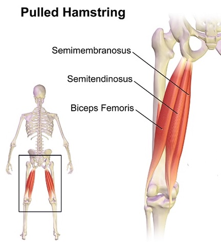
Posterior compartment of the thigh is another important fascial compartment. The muscles in posterior compartment of the thigh lengthen the thigh and stretch the leg. It comprises knee flexors and hip extensors, together known as hamstrings. The posterior compartment is enclosed within fascia and separated from anterior compartment by two bends of deep fascia, called medial intermuscular septum and lateral intermuscular septum. Moreover, the posterior compartment is homologous to anterior compartment of arm. This compartment is supplied by sciatic nerve that runs along the longitudinal axis. The main sciatic nerves are the common Peroneal nerve and the Tibial nerve. Also the arteries perforating this compartment arise from profunda femoris. Below are muscles of posterior fascial compartment of the thigh:

Biceps femoris
This muscle has two heads; one long head and the other short head. The long head originates from inferior and inward imprint of the back part of tuberosity of the ischium and lower end of sacrotuberous ligament whereas the short head originates from the lateral lip of linea aspera. The long head fibers form spindle shaped belly that passes implicitly downward and laterally through the sciatic nerve ending in aponeurosis. This aponeurosis then becomes constricted into tendon which then inserts into lateral side of fibula and a small portion into lateral condyle of tibia. Tendon then divides into two parts holding fibular collateral ligament of knee-joint. Right from the posterior border, a thin extension is given off to fascia of leg. Lateral hamstring is formed by the tendon of insertion of this muscle.
The short head is supplied by common fibular branch of sciatic nerve (L5, S2) whereas the long head is supplied by tibial branch of sciatic nerve (L5, S2). Also the vascular supply is carried by network of perforating branches of profunda femoris artery, inferior gluteal artery and popliteal artery. The main function of biceps femoris is flexion at the knee.
Muscles of posterior fascial compartment of the thigh Assignment Help is provided to you by the experienced and learned tutors of assignmenthelp.net at nominal price.
Semitendinosus
The long superficial muscle at the back of thigh is semitendinosus and is mainly present at the posterior and medial side of thigh. This muscle originates from the inferior and middle imprint on the tuberosity of ischium and from aponeurosis. It is fusiform in appearance ending in below the middle of thigh in a form of long round tendon. Then it curves around the medial condyle of tibia passing over medial collateral ligament of knee-joint. It is further separated by bursa, and is finally interleaved in the upper part of middle surface of body of the tibia.
Semitendinosus is supplied by tibial part of sciatic nerve. This muscle helps in flexion of leg at knee joint, extension of thigh at hip and rotation of thigh and leg.
Semimembranosus
This muscle originates from ischial tuberosity inserting into medial condyle, margin near to tibia, intercondylar fossa of femur and lateral condyle of femur along with ligament at the back of knee. The tendon of origin spreads into aponeurosis. From aponeurosis, muscle fibers arise that joins to form another aponeurosis that covers lower part of the posterior surface of the muscle, contracting into the tendon of insertion.
The insertion mainly takes place in the posterior medial aspect of the medial condyle of the tibia. It is wider, flatter and deeper than semitendinuous. The fibrous expansion is given off by tendon of insertion. One passes upward and laterally inserting into posterior lateral condyle of the femur, therefore forming oblique popliteal ligament of knee-joint. Likewise second goes downward to fascia covering the popliteus muscle. Likewise, collateral ligament of the joint and the fascia of the leg is joint by some fibers. This muscle is supplied by tibial part of sciatic nerve and helps in extension of hip joint and flexion of knee joint. Image reference: Commons.wikimedia.org
Want help with the Anatomical topics in Biology? submit assignment to get muscles of posterior fascial compartment of the thigh Assignment Help before the provided deadline.
| MUSCLE | ORIGIN | INSERTION | NERVE SUPPLY | NERVE ROOTS | ACTION |
| Biceps femoris | Ischial tuberosity, linea aspra, lateral supracondylar ridge of shaft of femur | Head of fibula | Tibial portion of sciatic nerve& Common peroneal nerve | L5,S1,2 | Flexes & laterally rotates leg at knee joint |
| Semitendinosus | Ischial tuberosity | Upper part of medial surface of shaft of tibia | Tibial portion of sciatic nerve | L5,S1,2 | Flexes & medially rotates leg at knee joint |
| Semimembranosus | Ischial tuberosity | Medial condyl of tibia | Tibial portion of sciatic nerve | L5,S1,2 | Flexes & medially rotates leg at knee joint |
Email Based Homework Help in Muscles Of Posterior Fascial Compartment Of The Thigh
To submit Muscles Of Posterior Fascial Compartment Of The Thigh assignment upload assignment.


